 Movie
Movie Controller

[English] 日本語
 Yorodumi
- PDB-5m6g: Crystal structure Glucan 1,4-beta-glucosidase from Saccharopolysp... -
+ Open data

- Basic information
| Entry | Database: PDB / ID: 5m6g | ||||||
|---|---|---|---|---|---|---|---|
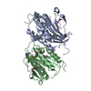
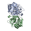
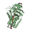
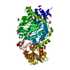
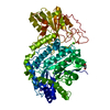
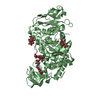
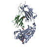
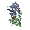
| Title | Crystal structure Glucan 1,4-beta-glucosidase from Saccharopolyspora erythraea | ||||||
 Components Components | Beta-glucosidase | ||||||
 Keywords Keywords | HYDROLASE / 2-DOMAIN FOLD | ||||||
| Function / homology |  Function and homology information Function and homology informationglucan 1,4-beta-glucosidase activity / glucan catabolic process / beta-glucosidase / metal ion binding Similarity search - Function | ||||||
| Biological species |  Saccharopolyspora erythraea D (bacteria) Saccharopolyspora erythraea D (bacteria) | ||||||
| Method |  X-RAY DIFFRACTION / SYNCHROTRON / MOLECULAR REPLACEMENT / Resolution: 1.829 Å X-RAY DIFFRACTION / SYNCHROTRON / MOLECULAR REPLACEMENT / Resolution: 1.829 Å | ||||||
 Authors Authors | Gabdulkhakov, A. / Tishchenko, S. / Lisov, A. / Leontievsky, A. | ||||||
 Citation Citation | Journal: To Be Published Title: Crystal structure Glucan 1,4-beta-glucosidase from Saccharopolyspora erythraea Authors: Gabdulkhakov, A. / Tishchenko, S. | ||||||
| History |
|
- Structure visualization
| Structure viewer | Molecule: MolmilJmol/JSmol |
|---|
- Downloads & links
-Download
| PDBx/mmCIF format | 5m6g.cif.gz | 131.5 KB | Display | PDBx/mmCIF format |
|---|---|---|---|---|
| PDB format | pdb5m6g.ent.gz | 98.3 KB | Display | PDB format |
| PDBx/mmJSON format | 5m6g.json.gz | Tree view | PDBx/mmJSON format | |
| Others |  Other downloads Other downloads |
-Validation report
| Arichive directory | https://data.pdbj.org/pub/pdb/validation_reports/m6/5m6gftp://data.pdbj.org/pub/pdb/validation_reports/m6/5m6g | HTTPS FTP |
|---|
-Related structure data
| Related structure data | 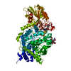 1ieqS S: Starting model for refinement |
|---|---|
| Similar structure data |


-Links
 PDBj
PDBj
- Assembly
| Deposited unit | 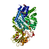
| ||||||||
|---|---|---|---|---|---|---|---|---|---|
| 1 |
| ||||||||
| Unit cell |
|
-Components
-Protein / Sugars , 2 types, 2 molecules A
| #1: Protein | Mass: 65379.684 Da / Num. of mol.: 1 Source method: isolated from a genetically manipulated source Source: (gene. exp.) Saccharopolyspora erythraea D (bacteria)Strain: ATCC 11635 / DSM 40517 / JCM 4748 / NBRC 13426 / NCIMB 8594 / NRRL 2338 Gene: celD, SACE_6502, A8924_6851 / Production host:  Komagataella pastoris GS115 (fungus) / References: UniProt: A4FNP6, glucan 1,4-beta-glucosidase Komagataella pastoris GS115 (fungus) / References: UniProt: A4FNP6, glucan 1,4-beta-glucosidase |
|---|---|
| #5: Sugar | ChemComp-SOR /  Type: D-saccharide / Mass: 182.172 Da / Num. of mol.: 1 Type: D-saccharide / Mass: 182.172 Da / Num. of mol.: 1Source method: isolated from a genetically manipulated source Formula: C6H14O6 |
-Non-polymers , 4 types, 288 molecules 


| #2: Chemical | ChemComp-MG /  Mass: 24.305 Da / Num. of mol.: 6 / Source method: obtained synthetically / Formula: Mg Mass: 24.305 Da / Num. of mol.: 6 / Source method: obtained synthetically / Formula: Mg#3: Chemical |  Mass: 92.094 Da / Num. of mol.: 3 / Source method: obtained synthetically / Formula: C3H8O3 Mass: 92.094 Da / Num. of mol.: 3 / Source method: obtained synthetically / Formula: C3H8O3#4: Chemical | ChemComp-PGE / |  Mass: 150.173 Da / Num. of mol.: 1 / Source method: obtained synthetically / Formula: C6H14O4 Mass: 150.173 Da / Num. of mol.: 1 / Source method: obtained synthetically / Formula: C6H14O4#6: Water | ChemComp-HOH / | Mass: 18.015 Da / Num. of mol.: 278 / Source method: isolated from a natural source / Formula: H2O |
|---|
-Details
| Has protein modification | Y |
|---|
-Experimental details
-Experiment
| Experiment | Method: X-RAY DIFFRACTION / Number of used crystals: 1 |
|---|
- Sample preparation
| Crystal | Density Matthews: 2.12 Å3/Da / Density % sol: 41.98 % |
|---|---|
| Crystal grow | Temperature: 295 K / Method: vapor diffusion, hanging drop / pH: 6.5 Details: 15% PEG 20K, 100mM MES Sodium Salt, 80 mM Mangan II Chloride |
-Data collection
| Diffraction | Mean temperature: 100 K |
|---|---|
| Diffraction source | Source: SYNCHROTRON / Site: PETRA III, DESY  / Beamline: P11 / Wavelength: 1.033121 Å / Beamline: P11 / Wavelength: 1.033121 Å |
| Detector | Type: DECTRIS PILATUS 6M / Detector: PIXEL / Date: Apr 17, 2016 |
| Radiation | Protocol: SINGLE WAVELENGTH / Monochromatic (M) / Laue (L): M / Scattering type: x-ray |
| Radiation wavelength | Wavelength: 1.033121 Å / Relative weight: 1 |
| Reflection | Resolution: 1.829→50 Å / Num. obs: 47457 / % possible obs: 95.1 % / Redundancy: 3.35 % / Net I/σ(I): 11.27 |
- Processing
| Software |
| ||||||||||||||||||||||||||||||||||||||||||||||||||||||||||||||||||||||||||||||||||||||||||||||||||||||||||||||||
|---|---|---|---|---|---|---|---|---|---|---|---|---|---|---|---|---|---|---|---|---|---|---|---|---|---|---|---|---|---|---|---|---|---|---|---|---|---|---|---|---|---|---|---|---|---|---|---|---|---|---|---|---|---|---|---|---|---|---|---|---|---|---|---|---|---|---|---|---|---|---|---|---|---|---|---|---|---|---|---|---|---|---|---|---|---|---|---|---|---|---|---|---|---|---|---|---|---|---|---|---|---|---|---|---|---|---|---|---|---|---|---|---|---|
| Refinement | Method to determine structure: MOLECULAR REPLACEMENT Starting model: 1ieq Resolution: 1.829→45.511 Å / SU ML: 0.19 / Cross valid method: FREE R-VALUE / σ(F): 1.35 / Phase error: 20.86
| ||||||||||||||||||||||||||||||||||||||||||||||||||||||||||||||||||||||||||||||||||||||||||||||||||||||||||||||||
| Solvent computation | Shrinkage radii: 0.9 Å / VDW probe radii: 1.11 Å | ||||||||||||||||||||||||||||||||||||||||||||||||||||||||||||||||||||||||||||||||||||||||||||||||||||||||||||||||
| Refinement step | Cycle: LAST / Resolution: 1.829→45.511 Å
| ||||||||||||||||||||||||||||||||||||||||||||||||||||||||||||||||||||||||||||||||||||||||||||||||||||||||||||||||
| Refine LS restraints |
| ||||||||||||||||||||||||||||||||||||||||||||||||||||||||||||||||||||||||||||||||||||||||||||||||||||||||||||||||
| LS refinement shell |
|
