[English] 日本語
Yorodumi
- PDB-5lyk: CRYSTAL STRUCTURE OF INTRACELLULAR B30.2 DOMAIN OF BTN3A1 BOUND T... -

+
Open data


ID or keywords:

Loading...

-
Basic information

Entry
Database: PDB / ID: 5lyk
Title
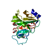
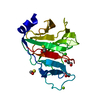
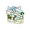
CRYSTAL STRUCTURE OF INTRACELLULAR B30.2 DOMAIN OF BTN3A1 BOUND TO CITRATE
ComponentsButyrophilin subfamily 3 member A1
KeywordsSIGNALING PROTEIN / B30.2 / butyrophilin
Function / homology
Function and homology information


Butyrophilin (BTN) family interactions / activated T cell proliferation / regulation of cytokine production / positive regulation of cytokine production / positive regulation of type II interferon production / T cell receptor signaling pathway / adaptive immune response / external side of plasma membrane / signaling receptor binding / plasma membrane
Similarity search - Function
Butyrophilin subfamily 3, PRY/SPRY domain / SPRY domain / SPRY-associated domain / SPRY-associated / PRY / Butyrophylin-like, SPRY domain / SPRY domain / B30.2/SPRY domain / B30.2/SPRY domain profile. / SPRY domain ...Butyrophilin subfamily 3, PRY/SPRY domain / SPRY domain / SPRY-associated domain / SPRY-associated / PRY / Butyrophylin-like, SPRY domain / SPRY domain / B30.2/SPRY domain / B30.2/SPRY domain profile. / SPRY domain / B30.2/SPRY domain superfamily / Domain in SPla and the RYanodine Receptor. / Immunoglobulin V-Type / Immunoglobulin V-set domain / Immunoglobulin V-set domain / Concanavalin A-like lectin/glucanase domain superfamily / Immunoglobulin subtype / Immunoglobulin / Jelly Rolls / Ig-like domain profile. / Immunoglobulin-like domain / Immunoglobulin-like domain superfamily / Immunoglobulin-like fold / Sandwich / Mainly Beta
Similarity search - Domain/homology
CITRATE ANION / Butyrophilin subfamily 3 member A1
Similarity search - Component
Biological speciesHomo sapiens (human)
MethodX-RAY DIFFRACTION / MOLECULAR REPLACEMENT / Resolution: 1.7 Å
AuthorsMohammed, F. / Baker, A.T. / Salim, M. / Willcox, B.E.
CitationJournal: ACS Chem. Biol. / Year: 2017
Title: BTN3A1 Discriminates gamma delta T Cell Phosphoantigens from Nonantigenic Small Molecules via a Conformational Sensor in Its B30.2 Domain.
Authors: Salim, M. / Knowles, T.J. / Baker, A.T. / Davey, M.S. / Jeeves, M. / Sridhar, P. / Wilkie, J. / Willcox, C.R. / Kadri, H. / Taher, T.E. / Vantourout, P. / Hayday, A. / Mehellou, Y. / Mohammed, F. / Willcox, B.E.
History
DepositionSep 28, 2016Deposition site: PDBE / Processing site: PDBE
Revision 1.0Sep 13, 2017Provider: repository / Type: Initial release
Revision 1.1Nov 1, 2017Group: Database references / Category: citation
Item: _citation.journal_volume / _citation.page_first ..._citation.journal_volume / _citation.page_first / _citation.page_last / _citation.title
Revision 1.2Jan 17, 2024Group: Advisory / Data collection ...Advisory / Data collection / Database references / Refinement description
Category: chem_comp_atom / chem_comp_bond ...chem_comp_atom / chem_comp_bond / database_2 / pdbx_initial_refinement_model / pdbx_unobs_or_zero_occ_atoms
Item: _database_2.pdbx_DOI / _database_2.pdbx_database_accession

-
Structure visualization

Structure viewerMolecule:
MolmilJmol/JSmol

Downloads & links

-
Assembly

Deposited unit
A: Butyrophilin subfamily 3 member A1
hetero molecules


Theoretical massNumber of molelcules
Total (without water)21,7413
Polymers21,4891
Non-polymers2512
Water3,315184
1


  • Idetical with deposited unit
  • defined by author&software
TypeNameSymmetry operationNumber
identity operation1_555x,y,z1
Buried area400 Å2
ΔGint6 kcal/mol
Surface area9400 Å2
MethodPISA
Unit cell
Length a, b, c (Å)45.500, 125.200, 39.200
Angle α, β, γ (deg.)90.000, 90.000, 90.000
Int Tables number18
Space group name H-MP21212

-
Components

#1: Protein Butyrophilin subfamily 3 member A1


Mass: 21489.451 Da / Num. of mol.: 1
Source method: isolated from a genetically manipulated source
Source: (gene. exp.) Homo sapiens (human) / Gene: BTN3A1, BTF5 / Plasmid: pET26 / Production host: ESCHERICHIA COLI BL21(DE3) (bacteria) / Variant (production host): pLysS / References: UniProt: O00481
#2: Chemical ChemComp-FLC / CITRATE ANION


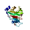
Mass: 189.100 Da / Num. of mol.: 1 / Source method: obtained synthetically / Formula: C6H5O7
#3: Chemical ChemComp-EDO / 1,2-ETHANEDIOL / ETHYLENE GLYCOL


Mass: 62.068 Da / Num. of mol.: 1 / Source method: obtained synthetically / Formula: C2H6O2
#4: Water ChemComp-HOH / water


Mass: 18.015 Da / Num. of mol.: 184 / Source method: isolated from a natural source / Formula: H2O

-
Experimental details

-
Experiment

ExperimentMethod: X-RAY DIFFRACTION / Number of used crystals: 1

-
Sample preparation

CrystalDensity Matthews: 2.6 Å3/Da / Density % sol: 52.65 %
Crystal growTemperature: 298 K / Method: vapor diffusion, hanging drop / pH: 7 / Details: 20% PEG 3350 and 0.2M Ammonium Citrate tribasic

-
Data collection

DiffractionMean temperature: 100 K
Diffraction sourceSource: ROTATING ANODE / Type: RIGAKU / Wavelength: 1.5417 Å
DetectorType: RIGAKU SATURN 944 / Detector: CCD / Date: Apr 14, 2016
RadiationProtocol: SINGLE WAVELENGTH / Monochromatic (M) / Laue (L): M / Scattering type: x-ray
Radiation wavelengthWavelength: 1.5417 Å / Relative weight: 1
ReflectionResolution: 1.7→63 Å / Num. obs: 24696 / % possible obs: 96.8 % / Observed criterion σ(I): -3 / Redundancy: 12 % / Biso Wilson estimate: 22.255 Å2 / CC1/2: 0.999 / Rmerge(I) obs: 0.058 / Net I/σ(I): 30.13
Reflection shell
Resolution (Å)Highest resolution (Å)Rmerge(I) obsMean I/σ(I) obsCC1/2Diffraction-ID% possible all
1.7-1.80.4734.060.861187
1.8-1.90.3136.880.953194.1
1.9-20.20610.410.985196.6
2-2.10.15214.550.99199.2
2.1-2.40.10923.410.9961100
2.4-2.70.07734.370.9981100
2.7-30.05847.420.9991100
3-3.50.04263.050.9991100
3.5-40.03478.4611100
4-50.0389.3411100
5-60.03287.3911100
6-100.03185.6711100
100.03179.790.998195.2

-
Processing

Software
NameVersionClassification
XSCALEdata scaling
REFMAC5.2.0019refinement
PDB_EXTRACT3.2data extraction
XDSdata reduction
MOLREPphasing
RefinementMethod to determine structure: MOLECULAR REPLACEMENT
Starting model: 4N7I
Resolution: 1.7→62.62 Å / Cor.coef. Fo:Fc: 0.95 / Cor.coef. Fo:Fc free: 0.94 / SU B: 4.374 / SU ML: 0.065 / SU R Cruickshank DPI: 0.146 / Cross valid method: THROUGHOUT / σ(F): 0 / ESU R: 0.146 / ESU R Free: 0.099 / Details: HYDROGENS HAVE BEEN ADDED IN THE RIDING POSITIONS
RfactorNum. reflection% reflectionSelection details
Rfree0.2089 1194 4.8 %RANDOM
Rwork0.181 ---
obs0.1824 23457 97.02 %-
Solvent computationIon probe radii: 0.8 Å / Shrinkage radii: 0.8 Å / VDW probe radii: 1.2 Å
Displacement parametersBiso max: 44.09 Å2 / Biso mean: 15.934 Å2 / Biso min: 2.95 Å2
Baniso -1Baniso -2Baniso -3
1--0.1 Å20 Å20 Å2
2--0.66 Å20 Å2
3----0.56 Å2
Refinement stepCycle: final / Resolution: 1.7→62.62 Å
ProteinNucleic acidLigandSolventTotal
Num. atoms1517 0 17 184 1718
Biso mean--20.79 25.41 -
Num. residues----187
Refine LS restraints
Refine-IDTypeDev idealDev ideal targetNumber
X-RAY DIFFRACTIONr_bond_refined_d0.0120.0221573
X-RAY DIFFRACTIONr_angle_refined_deg1.3091.9592142
X-RAY DIFFRACTIONr_dihedral_angle_1_deg5.8975186
X-RAY DIFFRACTIONr_dihedral_angle_2_deg32.50323.83673
X-RAY DIFFRACTIONr_dihedral_angle_3_deg11.27215253
X-RAY DIFFRACTIONr_dihedral_angle_4_deg10.409159
X-RAY DIFFRACTIONr_chiral_restr0.0920.2227
X-RAY DIFFRACTIONr_gen_planes_refined0.0050.021218
X-RAY DIFFRACTIONr_nbd_refined0.20.2668
X-RAY DIFFRACTIONr_nbtor_refined0.3060.21059
X-RAY DIFFRACTIONr_xyhbond_nbd_refined0.1180.2154
X-RAY DIFFRACTIONr_symmetry_vdw_refined0.1980.245
X-RAY DIFFRACTIONr_symmetry_hbond_refined0.1120.27
X-RAY DIFFRACTIONr_mcbond_it1.0261.5958
X-RAY DIFFRACTIONr_mcangle_it1.6421534
X-RAY DIFFRACTIONr_scbond_it2.2473707
X-RAY DIFFRACTIONr_scangle_it3.4224.5608
X-RAY DIFFRACTIONr_rigid_bond_restr1.38131665
X-RAY DIFFRACTIONr_sphericity_free4.1793184
X-RAY DIFFRACTIONr_sphericity_bonded2.01131529
LS refinement shellResolution: 1.701→1.745 Å / Total num. of bins used: 20
RfactorNum. reflection% reflection
Rfree0.314 73 -
Rwork0.243 1465 -
all-1538 -
obs--84.51 %

+
About Yorodumi

-
News

-
Feb 9, 2022. New format data for meta-information of EMDB entries

New format data for meta-information of EMDB entries

  • Version 3 of the EMDB header file is now the official format.
  • The previous official version 1.9 will be removed from the archive.

Related info.:EMDB header

External links:wwPDB to switch to version 3 of the EMDB data model

-
Aug 12, 2020. Covid-19 info

Covid-19 info

URL: https://pdbjlvh1.pdbj.org/emnavi/covid19.php

New page: Covid-19 featured information page in EM Navigator.

Related info.:Covid-19 info / Mar 5, 2020. Novel coronavirus structure data

+
Mar 5, 2020. Novel coronavirus structure data

Novel coronavirus structure data

Related info.:Yorodumi Speices / Aug 12, 2020. Covid-19 info

External links:COVID-19 featured content - PDBj / Molecule of the Month (242):Coronavirus Proteases

+
Jan 31, 2019. EMDB accession codes are about to change! (news from PDBe EMDB page)

EMDB accession codes are about to change! (news from PDBe EMDB page)

  • The allocation of 4 digits for EMDB accession codes will soon come to an end. Whilst these codes will remain in use, new EMDB accession codes will include an additional digit and will expand incrementally as the available range of codes is exhausted. The current 4-digit format prefixed with “EMD-” (i.e. EMD-XXXX) will advance to a 5-digit format (i.e. EMD-XXXXX), and so on. It is currently estimated that the 4-digit codes will be depleted around Spring 2019, at which point the 5-digit format will come into force.
  • The EM Navigator/Yorodumi systems omit the EMD- prefix.

Related info.:Q: What is EMD? / ID/Accession-code notation in Yorodumi/EM Navigator

External links:EMDB Accession Codes are Changing Soon! / Contact to PDBj

+
Jul 12, 2017. Major update of PDB

Major update of PDB

  • wwPDB released updated PDB data conforming to the new PDBx/mmCIF dictionary.
  • This is a major update changing the version number from 4 to 5, and with Remediation, in which all the entries are updated.
  • In this update, many items about electron microscopy experimental information are reorganized (e.g. em_software).
  • Now, EM Navigator and Yorodumi are based on the updated data.

External links:wwPDB Remediation / Enriched Model Files Conforming to OneDep Data Standards Now Available in the PDB FTP Archive

-
Yorodumi

Thousand views of thousand structures

  • Yorodumi is a browser for structure data from EMDB, PDB, SASBDB, etc.
  • This page is also the successor to EM Navigator detail page, and also detail information page/front-end page for Omokage search.
  • The word "yorodu" (or yorozu) is an old Japanese word meaning "ten thousand". "mi" (miru) is to see.

Related info.:EMDB / PDB / SASBDB / Comparison of 3 databanks / Yorodumi Search / Aug 31, 2016. New EM Navigator & Yorodumi / Yorodumi Papers / Jmol/JSmol / Function and homology information / Changes in new EM Navigator and Yorodumi

Read more