 Movie
Movie Controller

[English] 日本語
 Yorodumi
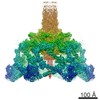
- PDB-5lye: Re-refined structure of the bacteriophage T4 short tail fibre PDB... -
+ Open data

- Basic information
| Entry | Database: PDB / ID: 5lye | |||||||||
|---|---|---|---|---|---|---|---|---|---|---|
| Title | Re-refined structure of the bacteriophage T4 short tail fibre PDB entry 1H6W containing 71 additionally identified residues | |||||||||
 Components Components | Gp12 | |||||||||
 Keywords Keywords | STRUCTURAL PROTEIN / GENE PRODUCT 12 / ADHESIN / FIBROUS PROTEIN | |||||||||
| Function / homology |  Function and homology information Function and homology information | |||||||||
| Biological species |  Enterobacteria phage T2 (virus) Enterobacteria phage T2 (virus) | |||||||||
| Method |  X-RAY DIFFRACTION / SYNCHROTRON / SIRAS / Resolution: 1.9 Å X-RAY DIFFRACTION / SYNCHROTRON / SIRAS / Resolution: 1.9 Å | |||||||||
 Authors Authors | van Raaij, M.J. / Taylor, N.M.I. / Leiman, P.G. | |||||||||
| Funding support |  Spain, 1items Spain, 1items
| |||||||||
 Citation Citation | Journal: Mol. Microbiol. / Year: 2018 Title: Contractile injection systems of bacteriophages and related systems. Authors: Taylor, N.M.I. / van Raaij, M.J. / Leiman, P.G. #1: Journal: Nature / Year: 2016Title: Structure of the T4 baseplate and its function in triggering sheath contraction. Authors: Nicholas M I Taylor / Nikolai S Prokhorov / Ricardo C Guerrero-Ferreira / Mikhail M Shneider / Christopher Browning / Kenneth N Goldie / Henning Stahlberg / Petr G Leiman /   Abstract: Several systems, including contractile tail bacteriophages, the type VI secretion system and R-type pyocins, use a multiprotein tubular apparatus to attach to and penetrate host cell membranes. This ...Several systems, including contractile tail bacteriophages, the type VI secretion system and R-type pyocins, use a multiprotein tubular apparatus to attach to and penetrate host cell membranes. This macromolecular machine resembles a stretched, coiled spring (or sheath) wound around a rigid tube with a spike-shaped protein at its tip. A baseplate structure, which is arguably the most complex part of this assembly, relays the contraction signal to the sheath. Here we present the atomic structure of the approximately 6-megadalton bacteriophage T4 baseplate in its pre- and post-host attachment states and explain the events that lead to sheath contraction in atomic detail. We establish the identity and function of a minimal set of components that is conserved in all contractile injection systems and show that the triggering mechanism is universally conserved. #2: Journal: J. Mol. Biol. / Year: 2001 Title: Crystal structure of a heat and protease-stable part of the bacteriophage T4 short tail fibre. Authors: van Raaij, M.J. / Schoehn, G. / Burda, M.R. / Miller, S. #3: Journal: Biol. Chem. / Year: 2001 Title: Identification and crystallisation of a heat- and protease-stable fragment of the bacteriophage T4 short tail fibre. Authors: van Raaij, M.J. / Schoehn, G. / Jaquinod, M. / Ashman, K. / Burda, M.R. / Miller, S. | |||||||||
| History |
|
- Structure visualization
| Structure viewer | Molecule: MolmilJmol/JSmol |
|---|
- Downloads & links
-Download
| PDBx/mmCIF format | 5lye.cif.gz | 66.7 KB | Display | PDBx/mmCIF format |
|---|---|---|---|---|
| PDB format | pdb5lye.ent.gz | 46.6 KB | Display | PDB format |
| PDBx/mmJSON format | 5lye.json.gz | Tree view | PDBx/mmJSON format | |
| Others |  Other downloads Other downloads |
-Validation report
| Arichive directory | https://data.pdbj.org/pub/pdb/validation_reports/ly/5lyeftp://data.pdbj.org/pub/pdb/validation_reports/ly/5lye | HTTPS FTP |
|---|
-Related structure data
| Related structure data | |
|---|---|
| Similar structure data |
-Links
 PDBj
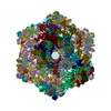
PDBj- Assembly
| Deposited unit | 
| |||||||||||||||
|---|---|---|---|---|---|---|---|---|---|---|---|---|---|---|---|---|
| 1 | 
| |||||||||||||||
| Unit cell |
| |||||||||||||||
| Components on special symmetry positions |
|
-Components
| #1: Protein | Mass: 33668.434 Da / Num. of mol.: 1 / Fragment: Heat and Protease-stable fragment Source method: isolated from a genetically manipulated source Source: (gene. exp.) Enterobacteria phage T2 (virus) / Gene: 12 / Production host:  | ||||||
|---|---|---|---|---|---|---|---|
| #2: Chemical |   Mass: 35.453 Da / Num. of mol.: 2 / Source method: obtained synthetically / Formula: Cl Mass: 35.453 Da / Num. of mol.: 2 / Source method: obtained synthetically / Formula: Cl#3: Chemical | ChemComp-SO4 / |   Mass: 96.063 Da / Num. of mol.: 1 / Source method: obtained synthetically / Formula: SO4 Mass: 96.063 Da / Num. of mol.: 1 / Source method: obtained synthetically / Formula: SO4#4: Water | ChemComp-HOH / |  Mass: 18.015 Da / Num. of mol.: 226 / Source method: isolated from a natural source / Formula: H2O Mass: 18.015 Da / Num. of mol.: 226 / Source method: isolated from a natural source / Formula: H2OSequence details | THE SEQUENCE OF THE SHORT TAIL FIBRE OF BACTERIOPH | |
-Experimental details
-Experiment
| Experiment | Method: X-RAY DIFFRACTION / Number of used crystals: 1 |
|---|
- Sample preparation
| Crystal | Density Matthews: 2.8 Å3/Da / Density % sol: 56 % / Description: Bars with hexagonal cross-section |
|---|---|
| Crystal grow | Temperature: 293 K / Method: vapor diffusion, sitting drop / pH: 5.6 Details: 15-25 % (V/V) 2-METHYLPROPANE-2-OL (TERTIARY BUTANOL), 100 MM SODIUM CITRATE PH 5.6 10% (V/V) GLYCEROL, 10 MM HEPES, 150 MM SODIUM CHLORIDE PH range: 5.6 |
-Data collection
| Diffraction | Mean temperature: 100 K |
|---|---|
| Diffraction source | Source: SYNCHROTRON / Site: ESRF  / Beamline: ID14-2 / Wavelength: 0.933 Å / Beamline: ID14-2 / Wavelength: 0.933 Å |
| Detector | Type: ADSC QUANTUM 4 / Detector: CCD / Date: Jun 1, 2000 |
| Radiation | Protocol: SINGLE WAVELENGTH / Monochromatic (M) / Laue (L): M / Scattering type: x-ray |
| Radiation wavelength | Wavelength: 0.933 Å / Relative weight: 1 |
| Reflection | Resolution: 1.9→19.8 Å / Num. obs: 28682 / % possible obs: 92.3 % / Redundancy: 7.3 % / Biso Wilson estimate: 16.12 Å2 / Rmerge(I) obs: 0.057 / Net I/σ(I): 8.5 |
| Reflection shell | Resolution: 1.9→2 Å / Redundancy: 5.3 % / Rmerge(I) obs: 0.154 / Mean I/σ(I) obs: 4.8 / % possible all: 68.7 |
- Processing
| Software |
| ||||||||||||||||||||||||||||||||||||||||||||||||||||||||||||||||||||||||||||||||||||||||||||||||||||||||||||||||||||||||||||||||||||||||||||||||||||||||||||||||||||||||||||||||||||||
|---|---|---|---|---|---|---|---|---|---|---|---|---|---|---|---|---|---|---|---|---|---|---|---|---|---|---|---|---|---|---|---|---|---|---|---|---|---|---|---|---|---|---|---|---|---|---|---|---|---|---|---|---|---|---|---|---|---|---|---|---|---|---|---|---|---|---|---|---|---|---|---|---|---|---|---|---|---|---|---|---|---|---|---|---|---|---|---|---|---|---|---|---|---|---|---|---|---|---|---|---|---|---|---|---|---|---|---|---|---|---|---|---|---|---|---|---|---|---|---|---|---|---|---|---|---|---|---|---|---|---|---|---|---|---|---|---|---|---|---|---|---|---|---|---|---|---|---|---|---|---|---|---|---|---|---|---|---|---|---|---|---|---|---|---|---|---|---|---|---|---|---|---|---|---|---|---|---|---|---|---|---|---|---|
| Refinement | Method to determine structure: SIRAS / Resolution: 1.9→19.8 Å / Cor.coef. Fo:Fc: 0.936 / Cor.coef. Fo:Fc free: 0.901 / SU B: 2.129 / SU ML: 0.064 / Cross valid method: THROUGHOUT / ESU R: 0.116 / ESU R Free: 0.115 / Details: HYDROGENS HAVE BEEN ADDED IN THE RIDING POSITIONS
| ||||||||||||||||||||||||||||||||||||||||||||||||||||||||||||||||||||||||||||||||||||||||||||||||||||||||||||||||||||||||||||||||||||||||||||||||||||||||||||||||||||||||||||||||||||||
| Solvent computation | Ion probe radii: 0.8 Å / Shrinkage radii: 0.8 Å / VDW probe radii: 1.2 Å | ||||||||||||||||||||||||||||||||||||||||||||||||||||||||||||||||||||||||||||||||||||||||||||||||||||||||||||||||||||||||||||||||||||||||||||||||||||||||||||||||||||||||||||||||||||||
| Displacement parameters | Biso mean: 37.983 Å2
| ||||||||||||||||||||||||||||||||||||||||||||||||||||||||||||||||||||||||||||||||||||||||||||||||||||||||||||||||||||||||||||||||||||||||||||||||||||||||||||||||||||||||||||||||||||||
| Refinement step | Cycle: LAST / Resolution: 1.9→19.8 Å
| ||||||||||||||||||||||||||||||||||||||||||||||||||||||||||||||||||||||||||||||||||||||||||||||||||||||||||||||||||||||||||||||||||||||||||||||||||||||||||||||||||||||||||||||||||||||
| Refine LS restraints |
|