 Movie
Movie Controller


+ Open data

- Basic information
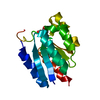
| Entry | Database: PDB / ID: 5lxu | ||||||
|---|---|---|---|---|---|---|---|
| Title | Structure of the DNA-binding domain of LUX ARRHYTHMO | ||||||
 Components Components |
| ||||||
 Keywords Keywords | DNA BINDING PROTEIN / DNA-binding / transcription factor / MYB domain / three helix bundle | ||||||
| Function / homology |  Function and homology information Function and homology informationpositive regulation of circadian rhythm / circadian rhythm / regulation of circadian rhythm / regulation of gene expression / transcription regulator complex / DNA-binding transcription factor activity / regulation of DNA-templated transcription / negative regulation of transcription by RNA polymerase II / DNA binding / nucleus Similarity search - Function | ||||||
| Biological species |  | ||||||
| Method |  X-RAY DIFFRACTION / SYNCHROTRON / SAD / Resolution: 2.14 Å X-RAY DIFFRACTION / SYNCHROTRON / SAD / Resolution: 2.14 Å | ||||||
 Authors Authors | Zubieta, C. / Nanao, M.H. | ||||||
| Funding support |  France, 1items France, 1items
| ||||||
 Citation Citation | Journal: To Be Published Title: Structure of the DNA-binding domain of LUX ARRHYTHMO Authors: Zubieta, C. / Silva, C.S. / Lai, X. / Wigge, P. / Nanao, M.H. / Nayak, A. | ||||||
| History |
|
- Structure visualization
| Structure viewer | Molecule: MolmilJmol/JSmol |
|---|
- Downloads & links
-Download
| PDBx/mmCIF format | 5lxu.cif.gz | 63.8 KB | Display | PDBx/mmCIF format |
|---|---|---|---|---|
| PDB format | pdb5lxu.ent.gz | 43.4 KB | Display | PDB format |
| PDBx/mmJSON format | 5lxu.json.gz | Tree view | PDBx/mmJSON format | |
| Others |  Other downloads Other downloads |
-Validation report
| Arichive directory | https://data.pdbj.org/pub/pdb/validation_reports/lx/5lxuftp://data.pdbj.org/pub/pdb/validation_reports/lx/5lxu | HTTPS FTP |
|---|
-Related structure data
| Similar structure data |
|---|


-Links
 PDBj
PDBj
- Assembly
| Deposited unit | 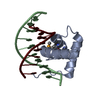
| ||||||||
|---|---|---|---|---|---|---|---|---|---|
| 1 |
| ||||||||
| Unit cell |
|
-Components
| #1: Protein | Mass: 6814.810 Da / Num. of mol.: 1 Source method: isolated from a genetically manipulated source Source: (gene. exp.)  |
|---|---|
| #2: DNA chain | Mass: 3035.003 Da / Num. of mol.: 1 / Source method: obtained synthetically / Source: (synth.) |
| #3: DNA chain | Mass: 3053.031 Da / Num. of mol.: 1 / Source method: obtained synthetically / Source: (synth.) |
| #4: Water | ChemComp-HOH /  Mass: 18.015 Da / Num. of mol.: 93 / Source method: isolated from a natural source / Formula: H2O Mass: 18.015 Da / Num. of mol.: 93 / Source method: isolated from a natural source / Formula: H2O |
| Has protein modification | Y |
-Experimental details
-Experiment
| Experiment | Method: X-RAY DIFFRACTION / Number of used crystals: 1 |
|---|
- Sample preparation
| Crystal | Density Matthews: 2.84 Å3/Da / Density % sol: 56.71 % |
|---|---|
| Crystal grow | Temperature: 293 K / Method: vapor diffusion, sitting drop / pH: 6.5 Details: 0.1M bis-tris propane, 20% PEG 3350, 0.2M sodium malonate |
-Data collection
| Diffraction | Mean temperature: 100 K | |||||||||||||||||||||||||||||||||||||||||||||||||||||||||||||||||||||||||||||
|---|---|---|---|---|---|---|---|---|---|---|---|---|---|---|---|---|---|---|---|---|---|---|---|---|---|---|---|---|---|---|---|---|---|---|---|---|---|---|---|---|---|---|---|---|---|---|---|---|---|---|---|---|---|---|---|---|---|---|---|---|---|---|---|---|---|---|---|---|---|---|---|---|---|---|---|---|---|---|
| Diffraction source | Source: SYNCHROTRON / Site: ESRF / Beamline: ID29 / Wavelength: 0.97886 Å | |||||||||||||||||||||||||||||||||||||||||||||||||||||||||||||||||||||||||||||
| Detector | Type: DECTRIS PILATUS 6M / Detector: PIXEL / Date: May 1, 2015 | |||||||||||||||||||||||||||||||||||||||||||||||||||||||||||||||||||||||||||||
| Radiation | Protocol: SINGLE WAVELENGTH / Monochromatic (M) / Laue (L): M / Scattering type: x-ray | |||||||||||||||||||||||||||||||||||||||||||||||||||||||||||||||||||||||||||||
| Radiation wavelength | Wavelength: 0.97886 Å / Relative weight: 1 | |||||||||||||||||||||||||||||||||||||||||||||||||||||||||||||||||||||||||||||
| Reflection | Resolution: 2.14→42 Å / Num. obs: 13640 / % possible obs: 86.7 % / Observed criterion σ(I): -3 / Redundancy: 1.47 % / Biso Wilson estimate: 57.22 Å2 / CC1/2: 0.999 / Rmerge(I) obs: 0.032 / Net I/σ(I): 13.48 | |||||||||||||||||||||||||||||||||||||||||||||||||||||||||||||||||||||||||||||
| Reflection shell | Diffraction-ID: 1
|
-Phasing
| Phasing | Method: SAD |
|---|
- Processing
| Software |
| ||||||||||||||||||||||||||||||||||||||||||||||||||||||||||||||||||||||||||||||||||||||||||||||||||||||||||||
|---|---|---|---|---|---|---|---|---|---|---|---|---|---|---|---|---|---|---|---|---|---|---|---|---|---|---|---|---|---|---|---|---|---|---|---|---|---|---|---|---|---|---|---|---|---|---|---|---|---|---|---|---|---|---|---|---|---|---|---|---|---|---|---|---|---|---|---|---|---|---|---|---|---|---|---|---|---|---|---|---|---|---|---|---|---|---|---|---|---|---|---|---|---|---|---|---|---|---|---|---|---|---|---|---|---|---|---|---|---|
| Refinement | Method to determine structure: SAD / Resolution: 2.14→41.68 Å / Cor.coef. Fo:Fc: 0.947 / Cor.coef. Fo:Fc free: 0.927 / Rfactor Rfree error: 0 / SU R Cruickshank DPI: 0.217 / Cross valid method: THROUGHOUT / σ(F): 0 / SU R Blow DPI: 0.248 / SU Rfree Blow DPI: 0.191 / SU Rfree Cruickshank DPI: 0.181
| ||||||||||||||||||||||||||||||||||||||||||||||||||||||||||||||||||||||||||||||||||||||||||||||||||||||||||||
| Displacement parameters | Biso max: 121.53 Å2 / Biso mean: 56.45 Å2 / Biso min: 34.83 Å2
| ||||||||||||||||||||||||||||||||||||||||||||||||||||||||||||||||||||||||||||||||||||||||||||||||||||||||||||
| Refine analyze | Luzzati coordinate error obs: 0.32 Å | ||||||||||||||||||||||||||||||||||||||||||||||||||||||||||||||||||||||||||||||||||||||||||||||||||||||||||||
| Refinement step | Cycle: final / Resolution: 2.14→41.68 Å
| ||||||||||||||||||||||||||||||||||||||||||||||||||||||||||||||||||||||||||||||||||||||||||||||||||||||||||||
| Refine LS restraints |
| ||||||||||||||||||||||||||||||||||||||||||||||||||||||||||||||||||||||||||||||||||||||||||||||||||||||||||||
| LS refinement shell | Resolution: 2.14→2.39 Å / Total num. of bins used: 5
| ||||||||||||||||||||||||||||||||||||||||||||||||||||||||||||||||||||||||||||||||||||||||||||||||||||||||||||
| Refinement TLS params. | Method: refined / Refine-ID: X-RAY DIFFRACTION
| ||||||||||||||||||||||||||||||||||||||||||||||||||||||||||||||||||||||||||||||||||||||||||||||||||||||||||||
| Refinement TLS group |
|
