 Movie
Movie Controller

[English] 日本語
 Yorodumi
- PDB-5lv9: Crystal structure of thermophilic tryptophan halogenase (Th-Hal) ... -
+ Open data

- Basic information
| Entry | Database: PDB / ID: 5lv9 | ||||||
|---|---|---|---|---|---|---|---|
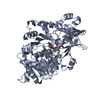
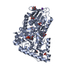
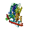
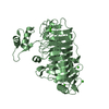
| Title | Crystal structure of thermophilic tryptophan halogenase (Th-Hal) enzyme from Streptomycin violaceusniger. | ||||||
 Components Components | thermophilic tryptophan halogenase | ||||||
 Keywords Keywords | HYDROLASE / thermophilic / flavin reductase / enzyme | ||||||
| Function / homology |  Function and homology information Function and homology information | ||||||
| Biological species |  Streptomyces violaceusniger (bacteria) Streptomyces violaceusniger (bacteria) | ||||||
| Method |  X-RAY DIFFRACTION / SYNCHROTRON / Resolution: 2.33 Å X-RAY DIFFRACTION / SYNCHROTRON / Resolution: 2.33 Å | ||||||
 Authors Authors | Dunstan, M.S. / Menon, B. | ||||||
| Funding support |  United Kingdom, 1items United Kingdom, 1items
| ||||||
 Citation Citation | Journal: Org.Biomol.Chem. / Year: 2016 Title: Structure and biocatalytic scope of thermophilic flavin-dependent halogenase and flavin reductase enzymes. Authors: Menon, B.R. / Latham, J. / Dunstan, M.S. / Brandenburger, E. / Klemstein, U. / Leys, D. / Karthikeyan, C. / Greaney, M.F. / Shepherd, S.A. / Micklefield, J. | ||||||
| History |
|
- Structure visualization
| Structure viewer | Molecule: MolmilJmol/JSmol |
|---|
- Downloads & links
-Download
| PDBx/mmCIF format | 5lv9.cif.gz | 414.8 KB | Display | PDBx/mmCIF format |
|---|---|---|---|---|
| PDB format | pdb5lv9.ent.gz | 344.2 KB | Display | PDB format |
| PDBx/mmJSON format | 5lv9.json.gz | Tree view | PDBx/mmJSON format | |
| Others |  Other downloads Other downloads |
-Validation report
| Arichive directory | https://data.pdbj.org/pub/pdb/validation_reports/lv/5lv9ftp://data.pdbj.org/pub/pdb/validation_reports/lv/5lv9 | HTTPS FTP |
|---|
-Related structure data


-Links
 PDBj
PDBj- Assembly
| Deposited unit | 
| ||||||||
|---|---|---|---|---|---|---|---|---|---|
| 1 | 
| ||||||||
| 2 | 
| ||||||||
| Unit cell |
|
-Components
| #1: Protein | Mass: 57969.336 Da / Num. of mol.: 2 Source method: isolated from a genetically manipulated source Source: (gene. exp.) Streptomyces violaceusniger (bacteria) / Production host: #2: Water | ChemComp-HOH / |  Mass: 18.015 Da / Num. of mol.: 377 / Source method: isolated from a natural source / Formula: H2O Mass: 18.015 Da / Num. of mol.: 377 / Source method: isolated from a natural source / Formula: H2O |
|---|
-Experimental details
-Experiment
| Experiment | Method: X-RAY DIFFRACTION / Number of used crystals: 1 |
|---|
- Sample preparation
| Crystal | Density Matthews: 2.68 Å3/Da / Density % sol: 54.09 % |
|---|---|
| Crystal grow | Temperature: 277 K / Method: vapor diffusion, sitting drop Details: 0.06 M divalents (0.3M Magnesium chloride hexahydrate; 0.3M Calcium chloride dehydrate) in 0.1 M buffer (1.0M imidazole + MES monohydrate acid buffer) at pH 6.5 made up with a 50% v/v ...Details: 0.06 M divalents (0.3M Magnesium chloride hexahydrate; 0.3M Calcium chloride dehydrate) in 0.1 M buffer (1.0M imidazole + MES monohydrate acid buffer) at pH 6.5 made up with a 50% v/v precipitant mix (that contained 25% v/v MPD; 25% PEG 1000; 25% w/v PEG 3350). |
-Data collection
| Diffraction | Mean temperature: 180 K |
|---|---|
| Diffraction source | Source: SYNCHROTRON / Site: Diamond / Beamline: I04 / Wavelength: 0.979 Å |
| Detector | Type: DECTRIS PILATUS3 S 6M / Detector: PIXEL / Date: Nov 28, 2014 |
| Radiation | Protocol: SINGLE WAVELENGTH / Monochromatic (M) / Laue (L): M / Scattering type: x-ray |
| Radiation wavelength | Wavelength: 0.979 Å / Relative weight: 1 |
| Reflection | Resolution: 2.33→57.7 Å / Num. obs: 51611 / % possible obs: 99.7 % / Redundancy: 9.9 % / Net I/σ(I): 11.7 |
- Processing
| Software |
| |||||||||||||||||||||||||||||||||||||||||||||||||||||||||||||||||||||||||||||||||||||||||||||||||||||||||||||||||||||||||||||||||||||||||||||||||||||||||||||||||||||||||||||||
|---|---|---|---|---|---|---|---|---|---|---|---|---|---|---|---|---|---|---|---|---|---|---|---|---|---|---|---|---|---|---|---|---|---|---|---|---|---|---|---|---|---|---|---|---|---|---|---|---|---|---|---|---|---|---|---|---|---|---|---|---|---|---|---|---|---|---|---|---|---|---|---|---|---|---|---|---|---|---|---|---|---|---|---|---|---|---|---|---|---|---|---|---|---|---|---|---|---|---|---|---|---|---|---|---|---|---|---|---|---|---|---|---|---|---|---|---|---|---|---|---|---|---|---|---|---|---|---|---|---|---|---|---|---|---|---|---|---|---|---|---|---|---|---|---|---|---|---|---|---|---|---|---|---|---|---|---|---|---|---|---|---|---|---|---|---|---|---|---|---|---|---|---|---|---|---|---|
| Refinement | Resolution: 2.33→56.493 Å / SU ML: 0.29 / Cross valid method: FREE R-VALUE / σ(F): 1.38 / Phase error: 24.2
| |||||||||||||||||||||||||||||||||||||||||||||||||||||||||||||||||||||||||||||||||||||||||||||||||||||||||||||||||||||||||||||||||||||||||||||||||||||||||||||||||||||||||||||||
| Solvent computation | Shrinkage radii: 0.9 Å / VDW probe radii: 1.11 Å | |||||||||||||||||||||||||||||||||||||||||||||||||||||||||||||||||||||||||||||||||||||||||||||||||||||||||||||||||||||||||||||||||||||||||||||||||||||||||||||||||||||||||||||||
| Displacement parameters | Biso max: 136.17 Å2 / Biso mean: 43.1279 Å2 / Biso min: 17.19 Å2 | |||||||||||||||||||||||||||||||||||||||||||||||||||||||||||||||||||||||||||||||||||||||||||||||||||||||||||||||||||||||||||||||||||||||||||||||||||||||||||||||||||||||||||||||
| Refinement step | Cycle: final / Resolution: 2.33→56.493 Å
| |||||||||||||||||||||||||||||||||||||||||||||||||||||||||||||||||||||||||||||||||||||||||||||||||||||||||||||||||||||||||||||||||||||||||||||||||||||||||||||||||||||||||||||||
| Refinement TLS params. | Method: refined / Refine-ID: X-RAY DIFFRACTION
| |||||||||||||||||||||||||||||||||||||||||||||||||||||||||||||||||||||||||||||||||||||||||||||||||||||||||||||||||||||||||||||||||||||||||||||||||||||||||||||||||||||||||||||||
| Refinement TLS group |
|
