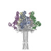
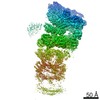
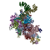
Journal: Nat Struct Mol Biol / Year: 2016 Title: Membrane insertion of a Tc toxin in near-atomic detail. Authors: Christos Gatsogiannis / Felipe Merino / Daniel Prumbaum / Daniel Roderer / Franziska Leidreiter / Dominic Meusch / Stefan Raunser / Abstract: Tc toxins from pathogenic bacteria use a special syringe-like mechanism to perforate the host cell membrane and inject a deadly enzyme into the host cytosol. The molecular mechanism of this unusual ...Tc toxins from pathogenic bacteria use a special syringe-like mechanism to perforate the host cell membrane and inject a deadly enzyme into the host cytosol. The molecular mechanism of this unusual injection system is poorly understood. Using electron cryomicroscopy, we determined the structure of TcdA1 from Photorhabdus luminescens embedded in lipid nanodiscs. In our structure, compared with the previous structure of TcdA1 in the prepore state, the transmembrane helices rearrange in the membrane and open the initially closed pore. However, the helices do not span the complete membrane; instead, the loops connecting the helices form the rim of the funnel. Lipid head groups reach into the space between the loops and consequently stabilize the pore conformation. The linker domain is folded and packed into a pocket formed by the other domains of the toxin, thereby considerably contributing to stabilization of the pore state.
Num. of particles selected: 30061 / Details: particles were picked manually
Symmetry
Point symmetry: C5 (5 fold cyclic)
3D reconstruction
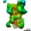
Resolution: 3.46 Å / Resolution method: FSC 0.143 CUT-OFF / Num. of particles: 13000 Details: The density was filtered to its local resolution, using localfilt (SPARX) and RESMAP Symmetry type: POINT
Atomic model building
Protocol: AB INITIO MODEL
Refinement
Highest resolution: 3.46 Å
Refine LS restraints
Refine-ID
Type
Dev ideal
Number
ELECTRONMICROSCOPY
f_bond_d
0.013
11395
ELECTRONMICROSCOPY
f_angle_d
1.127
15475
ELECTRONMICROSCOPY
f_dihedral_angle_d
11.213
9277
ELECTRONMICROSCOPY
f_chiral_restr
0.059
1708
ELECTRONMICROSCOPY
f_plane_restr
0.007
2018
+
About Yorodumi
-
News
-
Feb 9, 2022. New format data for meta-information of EMDB entries
New format data for meta-information of EMDB entries
Version 3 of the EMDB header file is now the official format.
The previous official version 1.9 will be removed from the archive.
In the structure databanks used in Yorodumi, some data are registered as the other names, "COVID-19 virus" and "2019-nCoV". Here are the details of the virus and the list of structure data.
Jan 31, 2019. EMDB accession codes are about to change! (news from PDBe EMDB page)
EMDB accession codes are about to change! (news from PDBe EMDB page)
The allocation of 4 digits for EMDB accession codes will soon come to an end. Whilst these codes will remain in use, new EMDB accession codes will include an additional digit and will expand incrementally as the available range of codes is exhausted. The current 4-digit format prefixed with “EMD-” (i.e. EMD-XXXX) will advance to a 5-digit format (i.e. EMD-XXXXX), and so on. It is currently estimated that the 4-digit codes will be depleted around Spring 2019, at which point the 5-digit format will come into force.
The EM Navigator/Yorodumi systems omit the EMD- prefix.
Related info.:Q: What is EMD? / ID/Accession-code notation in Yorodumi/EM Navigator
Yorodumi is a browser for structure data from EMDB, PDB, SASBDB, etc.
This page is also the successor to EM Navigator detail page, and also detail information page/front-end page for Omokage search.
The word "yorodu" (or yorozu) is an old Japanese word meaning "ten thousand". "mi" (miru) is to see.
Related info.:EMDB / PDB / SASBDB / Comparison of 3 databanks / Yorodumi Search / Aug 31, 2016. New EM Navigator & Yorodumi / Yorodumi Papers / Jmol/JSmol / Function and homology information / Changes in new EM Navigator and Yorodumi
 Movie
Movie Controller


 Open data
 Basic information
 Components
 Keywords
 Function and homology information
 Photorhabdus luminescens (bacteria)
 Authors
 Germany, 2items
  Citation
 Structure visualization
 Downloads & links
 Other downloads
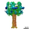
 PDBj
PDBj
 Assembly
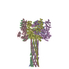
 Sample preparation
 Electron microscopy imaging
 FIELD EMISSION GUN / Accelerating voltage: 300 kV / Illumination mode: OTHER
 Processing