 Movie
Movie Controller


+ Open data

- Basic information
| Entry | Database: PDB / ID: 5l9w | ||||||
|---|---|---|---|---|---|---|---|
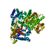
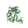
| Title | Crystal structure of the Apc core complex | ||||||
 Components Components | (Acetophenone carboxylase ...) x 4 | ||||||
 Keywords Keywords | LIGASE / Acetophenone carboxylase | ||||||
| Function / homology |  Function and homology information Function and homology informationacetophenone carboxylase / 5-oxoprolinase (ATP-hydrolyzing) activity / ligase activity / glutathione metabolic process / ATP binding / cytoplasm / cytosol Similarity search - Function | ||||||
| Biological species |  Aromatoleum aromaticum (bacteria) Aromatoleum aromaticum (bacteria) | ||||||
| Method |  X-RAY DIFFRACTION / SYNCHROTRON / SAD / Resolution: 2.9 Å X-RAY DIFFRACTION / SYNCHROTRON / SAD / Resolution: 2.9 Å | ||||||
 Authors Authors | Warkentin, E. / Weidenweber, S. / Ermler, U. | ||||||
 Citation Citation | Journal: Sci Rep / Year: 2017 Title: Structure of the acetophenone carboxylase core complex: prototype of a new class of ATP-dependent carboxylases/hydrolases. Authors: Weidenweber, S. / Schuhle, K. / Demmer, U. / Warkentin, E. / Ermler, U. / Heider, J. #1: Journal: To Be PublishedTitle: A new secondary structure in proteins: The G column, a bundle of glycine-rich type II polyproline helices Authors: Warkentin, E. / Weidenweber, S. / Schuehle, K. / Demmer, U. / Heider, J. / Ermler, U. | ||||||
| History |
|
- Structure visualization
| Structure viewer | Molecule: MolmilJmol/JSmol |
|---|
- Downloads & links
-Download
| PDBx/mmCIF format | 5l9w.cif.gz | 854.9 KB | Display | PDBx/mmCIF format |
|---|---|---|---|---|
| PDB format | pdb5l9w.ent.gz | 712.2 KB | Display | PDB format |
| PDBx/mmJSON format | 5l9w.json.gz | Tree view | PDBx/mmJSON format | |
| Others |  Other downloads Other downloads |
-Validation report
| Arichive directory | https://data.pdbj.org/pub/pdb/validation_reports/l9/5l9wftp://data.pdbj.org/pub/pdb/validation_reports/l9/5l9w | HTTPS FTP |
|---|
-Related structure data
| Similar structure data |
|---|


-Links
 PDBj
PDBj


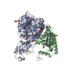
- Assembly
| Deposited unit | 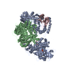
| ||||||||
|---|---|---|---|---|---|---|---|---|---|
| 1 | 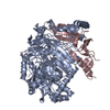
| ||||||||
| 2 | 
| ||||||||
| Unit cell |
|
-Components
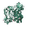
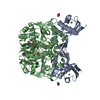
-Acetophenone carboxylase ... , 4 types, 4 molecules ABbC
| #1: Protein | Mass: 75484.609 Da / Num. of mol.: 1 / Source method: isolated from a natural source Source: (natural) Aromatoleum aromaticum (strain EbN1) (bacteria)Strain: EbN1 / References: UniProt: Q5P5G5, acetophenone carboxylase |
|---|---|
| #2: Protein | Mass: 80431.875 Da / Num. of mol.: 1 / Source method: isolated from a natural source Source: (natural) Aromatoleum aromaticum (strain EbN1) (bacteria)Strain: EbN1 / References: UniProt: Q5P5G4, acetophenone carboxylase |
| #3: Protein | Mass: 69935.141 Da / Num. of mol.: 1 / Source method: isolated from a natural source Source: (natural) Aromatoleum aromaticum (strain EbN1) (bacteria)Strain: EbN1 / References: UniProt: Q5P5G2, acetophenone carboxylase |
| #4: Protein | Mass: 15015.123 Da / Num. of mol.: 1 / Source method: isolated from a natural source Source: (natural) Aromatoleum aromaticum (strain EbN1) (bacteria)Strain: EbN1 / References: UniProt: Q5P5G3, acetophenone carboxylase |
-Non-polymers , 5 types, 23 molecules 


| #5: Chemical | ChemComp-HG /  Mass: 200.590 Da / Num. of mol.: 4 / Source method: obtained synthetically / Formula: Hg Mass: 200.590 Da / Num. of mol.: 4 / Source method: obtained synthetically / Formula: Hg#6: Chemical | ChemComp-K / |  Mass: 39.098 Da / Num. of mol.: 1 / Source method: obtained synthetically / Formula: K Mass: 39.098 Da / Num. of mol.: 1 / Source method: obtained synthetically / Formula: K#7: Chemical |  Mass: 238.305 Da / Num. of mol.: 3 / Source method: isolated from a natural source / Formula: C8H18N2O4S / Comment: pH buffer*YM Mass: 238.305 Da / Num. of mol.: 3 / Source method: isolated from a natural source / Formula: C8H18N2O4S / Comment: pH buffer*YM#8: Chemical | ChemComp-ADP / |  Mass: 427.201 Da / Num. of mol.: 1 / Source method: obtained synthetically / Formula: C10H15N5O10P2 / Comment: ADP, energy-carrying molecule*YM Mass: 427.201 Da / Num. of mol.: 1 / Source method: obtained synthetically / Formula: C10H15N5O10P2 / Comment: ADP, energy-carrying molecule*YM#9: Water | ChemComp-HOH / | Mass: 18.015 Da / Num. of mol.: 14 / Source method: isolated from a natural source / Formula: H2O |
|---|
-Details
| Has protein modification | Y |
|---|
-Experimental details
-Experiment
| Experiment | Method: X-RAY DIFFRACTION / Number of used crystals: 1 |
|---|
- Sample preparation
| Crystal | Density Matthews: 5.81 Å3/Da / Density % sol: 78.84 % |
|---|---|
| Crystal grow | Temperature: 277 K / Method: vapor diffusion, sitting drop / pH: 7.5 / Details: 34 - 50 % (v/v) PEE, 0.1 M Hepes |
-Data collection
| Diffraction | Mean temperature: 100 K |
|---|---|
| Diffraction source | Source: SYNCHROTRON / Site: SLS  / Beamline: X10SA / Wavelength: 1.008 Å / Beamline: X10SA / Wavelength: 1.008 Å |
| Detector | Type: DECTRIS PILATUS 6M-F / Detector: PIXEL / Date: Nov 11, 2013 |
| Radiation | Protocol: SINGLE WAVELENGTH / Monochromatic (M) / Laue (L): M / Scattering type: x-ray |
| Radiation wavelength | Wavelength: 1.008 Å / Relative weight: 1 |
| Reflection | Resolution: 2.9→50 Å / Num. obs: 121963 / % possible obs: 96.4 % / Redundancy: 14 % / Rrim(I) all: 0.117 / Net I/σ(I): 25 |
| Reflection shell | Resolution: 2.9→3.1 Å / Redundancy: 14.4 % / Mean I/σ(I) obs: 1.7 / Rrim(I) all: 2.42 / % possible all: 90.5 |
- Processing
| Software |
| |||||||||||||||||||||||||||||||||||||||||||||||||||||||||||||||||||||||||||||||||||||||||||||||||||||||||||||||||||||||||||||||||||||||||||||||||||||||||||||||||||||||||||||||||||||||||||||||||||||||||||||||||||||||||
|---|---|---|---|---|---|---|---|---|---|---|---|---|---|---|---|---|---|---|---|---|---|---|---|---|---|---|---|---|---|---|---|---|---|---|---|---|---|---|---|---|---|---|---|---|---|---|---|---|---|---|---|---|---|---|---|---|---|---|---|---|---|---|---|---|---|---|---|---|---|---|---|---|---|---|---|---|---|---|---|---|---|---|---|---|---|---|---|---|---|---|---|---|---|---|---|---|---|---|---|---|---|---|---|---|---|---|---|---|---|---|---|---|---|---|---|---|---|---|---|---|---|---|---|---|---|---|---|---|---|---|---|---|---|---|---|---|---|---|---|---|---|---|---|---|---|---|---|---|---|---|---|---|---|---|---|---|---|---|---|---|---|---|---|---|---|---|---|---|---|---|---|---|---|---|---|---|---|---|---|---|---|---|---|---|---|---|---|---|---|---|---|---|---|---|---|---|---|---|---|---|---|---|---|---|---|---|---|---|---|---|---|---|---|---|---|---|---|---|
| Refinement | Method to determine structure: SAD / Resolution: 2.9→19.971 Å / SU ML: 0.46 / Cross valid method: THROUGHOUT / σ(F): 1.35 / Phase error: 29.62
| |||||||||||||||||||||||||||||||||||||||||||||||||||||||||||||||||||||||||||||||||||||||||||||||||||||||||||||||||||||||||||||||||||||||||||||||||||||||||||||||||||||||||||||||||||||||||||||||||||||||||||||||||||||||||
| Solvent computation | Shrinkage radii: 0.9 Å / VDW probe radii: 1.11 Å | |||||||||||||||||||||||||||||||||||||||||||||||||||||||||||||||||||||||||||||||||||||||||||||||||||||||||||||||||||||||||||||||||||||||||||||||||||||||||||||||||||||||||||||||||||||||||||||||||||||||||||||||||||||||||
| Refinement step | Cycle: LAST / Resolution: 2.9→19.971 Å
| |||||||||||||||||||||||||||||||||||||||||||||||||||||||||||||||||||||||||||||||||||||||||||||||||||||||||||||||||||||||||||||||||||||||||||||||||||||||||||||||||||||||||||||||||||||||||||||||||||||||||||||||||||||||||
| Refine LS restraints |
| |||||||||||||||||||||||||||||||||||||||||||||||||||||||||||||||||||||||||||||||||||||||||||||||||||||||||||||||||||||||||||||||||||||||||||||||||||||||||||||||||||||||||||||||||||||||||||||||||||||||||||||||||||||||||
| LS refinement shell |
| |||||||||||||||||||||||||||||||||||||||||||||||||||||||||||||||||||||||||||||||||||||||||||||||||||||||||||||||||||||||||||||||||||||||||||||||||||||||||||||||||||||||||||||||||||||||||||||||||||||||||||||||||||||||||
| Refinement TLS params. | Method: refined / Refine-ID: X-RAY DIFFRACTION
| |||||||||||||||||||||||||||||||||||||||||||||||||||||||||||||||||||||||||||||||||||||||||||||||||||||||||||||||||||||||||||||||||||||||||||||||||||||||||||||||||||||||||||||||||||||||||||||||||||||||||||||||||||||||||
| Refinement TLS group |
|
