| Entry | Database: PDB / ID: 5ktk
|
|---|
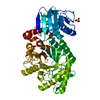
| Title | Ketoreductase from module 3 of the bacillaene synthase from Bacillus subtilis 168 |
|---|
 Components Components | Polyketide synthase PksJ |
|---|
 Keywords Keywords | OXIDOREDUCTASE / Bacillaene polyketide ketoreductase |
|---|
| Function / homology |  Function and homology information Function and homology information
DIM/DIP cell wall layer assembly / secondary metabolite biosynthetic process / fatty acid synthase activity / ligase activity / phosphopantetheine binding / 3-oxoacyl-[acyl-carrier-protein] synthase activity / antibiotic biosynthetic process / fatty acid biosynthetic process / cytoplasmSimilarity search - Function : / RhiE-like, KS-MAT linker domain / : / Polyketide synthase dehydratase N-terminal domain / Condensation domain / Condensation domain / Amino acid adenylation domain / Polyketide synthase, dehydratase domain / PKS_DH / PKS_PP_betabranch ...: / RhiE-like, KS-MAT linker domain / : / Polyketide synthase dehydratase N-terminal domain / Condensation domain / Condensation domain / Amino acid adenylation domain / Polyketide synthase, dehydratase domain / PKS_DH / PKS_PP_betabranch / : / Polyketide synthase dehydratase domain / : / Polyketide and metazoan fatty acid synthase dehydratase (PKS/mFAS DH) domain profile. / Polyketide synthase, dehydratase domain superfamily / Polyketide synthase, ketoreductase domain / KR domain / ANL, N-terminal domain / : / AMP-binding enzyme C-terminal domain / AMP-binding enzyme, C-terminal domain / AMP-binding, conserved site / Putative AMP-binding domain signature. / Chloramphenicol acetyltransferase-like domain superfamily / AMP-dependent synthetase/ligase / AMP-binding enzyme / Polyketide synthase, phosphopantetheine-binding domain / Phosphopantetheine attachment site / PKS_KR / Beta-ketoacyl synthase / Beta-ketoacyl synthase, active site / Ketosynthase family 3 (KS3) active site signature. / AMP-binding enzyme, C-terminal domain superfamily / Ketosynthase family 3 (KS3) domain profile. / Beta-ketoacyl synthase, N-terminal / Beta-ketoacyl synthase, C-terminal / Polyketide synthase, beta-ketoacyl synthase domain / Beta-ketoacyl synthase, N-terminal domain / Beta-ketoacyl synthase, C-terminal domain / Thiolase-like / Phosphopantetheine attachment site / Phosphopantetheine attachment site. / Phosphopantetheine attachment site / ACP-like superfamily / Carrier protein (CP) domain profile. / Phosphopantetheine binding ACP domain / NAD(P)-binding Rossmann-like Domain / NAD(P)-binding domain superfamily / Rossmann fold / 3-Layer(aba) Sandwich / Alpha BetaSimilarity search - Domain/homology |
|---|
| Biological species |   Bacillus subtilis (bacteria) Bacillus subtilis (bacteria) |
|---|
| Method |  X-RAY DIFFRACTION / SYNCHROTRON / MOLECULAR REPLACEMENT / Resolution: 1.98 Å X-RAY DIFFRACTION / SYNCHROTRON / MOLECULAR REPLACEMENT / Resolution: 1.98 Å |
|---|
 Authors Authors | Wagner, D.T. / Gay, D.C. / Keatinge-Clay, A.T. |
|---|
| Funding support |  United States, 1items United States, 1items | Organization | Grant number | Country |
|---|
| National Institutes of Health/National Institute of General Medical Sciences (NIH/NIGMS) | 2616061650 | United States |
|
|---|
 Citation Citation | Journal: To Be Published
Title: Ketoreductase from module 3 of the bacillaene synthase from Bacillus subtilis 168
Authors: Wagner, D.T. / Gay, D.C. / Keatinge-Clay, A.T. |
|---|
| History | | Deposition | Jul 11, 2016 | Deposition site: RCSB / Processing site: RCSB |
|---|
| Revision 1.0 | Sep 7, 2016 | Provider: repository / Type: Initial release |
|---|
| Revision 1.1 | Sep 20, 2017 | Group: Author supporting evidence / Derived calculations / Category: pdbx_audit_support / pdbx_struct_oper_list
Item: _pdbx_audit_support.funding_organization / _pdbx_struct_oper_list.symmetry_operation |
|---|
| Revision 1.2 | Dec 25, 2019 | Group: Author supporting evidence / Category: pdbx_audit_support / Item: _pdbx_audit_support.funding_organization |
|---|
| Revision 1.3 | Oct 4, 2023 | Group: Data collection / Database references / Refinement description
Category: chem_comp_atom / chem_comp_bond ...chem_comp_atom / chem_comp_bond / database_2 / pdbx_initial_refinement_model
Item: _database_2.pdbx_DOI / _database_2.pdbx_database_accession |
|---|
|
|---|
 Movie
Movie Controller

 Yorodumi
 Open data
 Basic information
 Structure visualization
 Downloads & links
 Other downloads

 PDBj
PDBj


 Assembly

 Mass: 745.421 Da / Num. of mol.: 1 / Source method: obtained synthetically / Formula: C21H30N7O17P3
 Mass: 18.015 Da / Num. of mol.: 155 / Source method: isolated from a natural source / Formula: H2O
 Sample preparation
 Processing