 Movie
Movie Controller


+ Open data

- Basic information
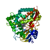
| Entry | Database: PDB / ID: 5kt1 | ||||||
|---|---|---|---|---|---|---|---|
| Title | The crystal structure of 4-methylthiobenzoate-bound CYP199A4 | ||||||
 Components Components | Cytochrome P450 | ||||||
 Keywords Keywords | OXIDOREDUCTASE / P450 / Substrate | ||||||
| Function / homology |  Function and homology information Function and homology informationoxidoreductase activity, acting on paired donors, with incorporation or reduction of molecular oxygen / monooxygenase activity / iron ion binding / heme binding Similarity search - Function | ||||||
| Biological species |  Rhodopseudomonas palustris (phototrophic) Rhodopseudomonas palustris (phototrophic) | ||||||
| Method |  X-RAY DIFFRACTION / SYNCHROTRON / MOLECULAR REPLACEMENT / molecular replacement / Resolution: 2.015 Å X-RAY DIFFRACTION / SYNCHROTRON / MOLECULAR REPLACEMENT / molecular replacement / Resolution: 2.015 Å | ||||||
 Authors Authors | Coleman, T. / Bruning, J.B. / Bell, S.G. | ||||||
| Funding support |  Australia, 1items Australia, 1items
| ||||||
 Citation Citation | Journal: To Be Published Title: The crystal structure of 4-methylthiobenzoate-bound CYP199A4 Authors: Coleman, T. / Bruning, J.B. / Bell, S.G. | ||||||
| History |
|
- Structure visualization
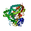
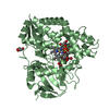
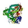
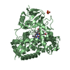
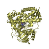
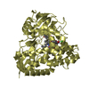
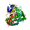
| Structure viewer | Molecule: MolmilJmol/JSmol |
|---|
- Downloads & links
-Download
| PDBx/mmCIF format | 5kt1.cif.gz | 103 KB | Display | PDBx/mmCIF format |
|---|---|---|---|---|
| PDB format | pdb5kt1.ent.gz | 73.9 KB | Display | PDB format |
| PDBx/mmJSON format | 5kt1.json.gz | Tree view | PDBx/mmJSON format | |
| Others |  Other downloads Other downloads |
-Validation report
| Arichive directory | https://data.pdbj.org/pub/pdb/validation_reports/kt/5kt1ftp://data.pdbj.org/pub/pdb/validation_reports/kt/5kt1 | HTTPS FTP |
|---|
-Related structure data
| Related structure data | 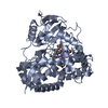 4do1S S: Starting model for refinement |
|---|---|
| Similar structure data |


-Links
 PDBj
PDBj
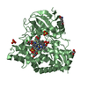
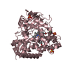
- Assembly
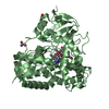
| Deposited unit | 
| ||||||||
|---|---|---|---|---|---|---|---|---|---|
| 1 |
| ||||||||
| Unit cell |
|
-Components
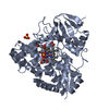
-Protein , 1 types, 1 molecules A
| #1: Protein | Mass: 42898.660 Da / Num. of mol.: 1 Source method: isolated from a genetically manipulated source Source: (gene. exp.) Rhodopseudomonas palustris (strain HaA2) (phototrophic)Strain: HaA2 / Gene: RPB_3613 / Production host: |
|---|
-Non-polymers , 5 types, 370 molecules 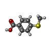


| #2: Chemical | ChemComp-4MI /  Mass: 168.213 Da / Num. of mol.: 1 / Source method: obtained synthetically / Formula: C8H8O2S Mass: 168.213 Da / Num. of mol.: 1 / Source method: obtained synthetically / Formula: C8H8O2S |
|---|---|
| #3: Chemical | ChemComp-HEM /  Mass: 616.487 Da / Num. of mol.: 1 / Source method: obtained synthetically / Formula: C34H32FeN4O4 Mass: 616.487 Da / Num. of mol.: 1 / Source method: obtained synthetically / Formula: C34H32FeN4O4 |
| #4: Chemical | ChemComp-CL /  Mass: 35.453 Da / Num. of mol.: 1 / Source method: obtained synthetically / Formula: Cl Mass: 35.453 Da / Num. of mol.: 1 / Source method: obtained synthetically / Formula: Cl |
| #5: Chemical | ChemComp-GOL /  Mass: 92.094 Da / Num. of mol.: 1 / Source method: obtained synthetically / Formula: C3H8O3 Mass: 92.094 Da / Num. of mol.: 1 / Source method: obtained synthetically / Formula: C3H8O3 |
| #6: Water | ChemComp-HOH / Mass: 18.015 Da / Num. of mol.: 366 / Source method: isolated from a natural source / Formula: H2O |
-Experimental details
-Experiment
| Experiment | Method: X-RAY DIFFRACTION / Number of used crystals: 1 |
|---|
- Sample preparation
| Crystal | Density Matthews: 2.08 Å3/Da / Density % sol: 40.84 % Description: rectangular plate - approx dimensions 300 um by 150 um by 20 um |
|---|---|
| Crystal grow | Temperature: 289.15 K / Method: vapor diffusion, hanging drop / pH: 5.5 Details: 0.2M Magnesium acetate PEG 3350 - 23% w/v Bis-Tris 0.1M - pH 5.5 PH range: 5.25-6 / Temp details: 16 celsius |
-Data collection
| Diffraction | Mean temperature: 100 K | ||||||||||||||||||
|---|---|---|---|---|---|---|---|---|---|---|---|---|---|---|---|---|---|---|---|
| Diffraction source | Source: SYNCHROTRON / Site: Australian Synchrotron / Beamline: MX2 / Wavelength: 0.9537 Å | ||||||||||||||||||
| Detector | Type: ADSC QUANTUM 315r / Detector: CCD / Date: Jun 21, 2016 | ||||||||||||||||||
| Radiation | Protocol: SINGLE WAVELENGTH / Monochromatic (M) / Laue (L): M / Scattering type: x-ray | ||||||||||||||||||
| Radiation wavelength | Wavelength: 0.9537 Å / Relative weight: 1 | ||||||||||||||||||
| Reflection | Resolution: 2.01→44.05 Å / Num. obs: 23479 / % possible obs: 99.7 % / Redundancy: 7.1 % / CC1/2: 0.984 / Rmerge(I) obs: 0.297 / Rpim(I) all: 0.119 / Rrim(I) all: 0.32 / Net I/σ(I): 7.2 / Num. measured all: 167452 / Scaling rejects: 6 | ||||||||||||||||||
| Reflection shell |
|
-Phasing
| Phasing | Method: molecular replacement | |||||||||
|---|---|---|---|---|---|---|---|---|---|---|
| Phasing MR | Model details: Phaser MODE: MR_AUTO
|
- Processing
| Software |
| ||||||||||||||||||||||||||||||||||||||||||||||||||||||||||||||||||||||
|---|---|---|---|---|---|---|---|---|---|---|---|---|---|---|---|---|---|---|---|---|---|---|---|---|---|---|---|---|---|---|---|---|---|---|---|---|---|---|---|---|---|---|---|---|---|---|---|---|---|---|---|---|---|---|---|---|---|---|---|---|---|---|---|---|---|---|---|---|---|---|---|
| Refinement | Method to determine structure: MOLECULAR REPLACEMENT Starting model: PDB: 4DO1 Resolution: 2.015→44.047 Å / SU ML: 0.23 / Cross valid method: FREE R-VALUE / σ(F): 1.35 / Phase error: 22.42
| ||||||||||||||||||||||||||||||||||||||||||||||||||||||||||||||||||||||
| Solvent computation | Shrinkage radii: 0.9 Å / VDW probe radii: 1.11 Å | ||||||||||||||||||||||||||||||||||||||||||||||||||||||||||||||||||||||
| Displacement parameters | Biso max: 74.7 Å2 / Biso mean: 18.5446 Å2 / Biso min: 4.88 Å2 | ||||||||||||||||||||||||||||||||||||||||||||||||||||||||||||||||||||||
| Refinement step | Cycle: final / Resolution: 2.015→44.047 Å
| ||||||||||||||||||||||||||||||||||||||||||||||||||||||||||||||||||||||
| Refine LS restraints |
| ||||||||||||||||||||||||||||||||||||||||||||||||||||||||||||||||||||||
| LS refinement shell | Refine-ID: X-RAY DIFFRACTION / Total num. of bins used: 9
|
