 Movie
Movie Controller

[English] 日本語
 Yorodumi
- PDB-5knk: Lipid A secondary acyltransferase LpxM from Acinetobacter baumann... -
+ Open data

- Basic information
| Entry | Database: PDB / ID: 5knk | ||||||
|---|---|---|---|---|---|---|---|
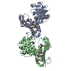
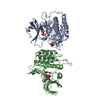
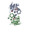
| Title | Lipid A secondary acyltransferase LpxM from Acinetobacter baumannii with catalytic residue substitution (E127A) | ||||||
 Components Components | Lipid A biosynthesis lauroyl acyltransferase | ||||||
 Keywords Keywords | TRANSFERASE / LpxM MsbB WaaN / Acyl Transferase / Lauryl Transferase | ||||||
| Function / homology | Bacterial lipid A biosynthesis acyltransferase / Bacterial lipid A biosynthesis acyltransferase / glycolipid biosynthetic process / acyltransferase activity / plasma membrane / UNDECANOIC ACID / Lipid A biosynthesis lauroyl acyltransferase Function and homology information Function and homology information | ||||||
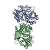
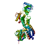
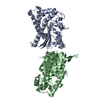
| Biological species |  Acinetobacter baumannii NIPH 410 (bacteria) Acinetobacter baumannii NIPH 410 (bacteria) | ||||||
| Method |  X-RAY DIFFRACTION / SYNCHROTRON / MOLECULAR REPLACEMENT / Resolution: 1.9 Å X-RAY DIFFRACTION / SYNCHROTRON / MOLECULAR REPLACEMENT / Resolution: 1.9 Å | ||||||
 Authors Authors | Dovala, D.L. / Hu, Q. / Metzger IV, L.E. | ||||||
 Citation Citation | Journal: Proc.Natl.Acad.Sci.USA / Year: 2016 Title: Structure-guided enzymology of the lipid A acyltransferase LpxM reveals a dual activity mechanism. Authors: Dovala, D. / Rath, C.M. / Hu, Q. / Sawyer, W.S. / Shia, S. / Elling, R.A. / Knapp, M.S. / Metzger, L.E. | ||||||
| History |
|
- Structure visualization
| Structure viewer | Molecule: MolmilJmol/JSmol |
|---|
- Downloads & links
-Download
| PDBx/mmCIF format | 5knk.cif.gz | 89 KB | Display | PDBx/mmCIF format |
|---|---|---|---|---|
| PDB format | pdb5knk.ent.gz | 64.3 KB | Display | PDB format |
| PDBx/mmJSON format | 5knk.json.gz | Tree view | PDBx/mmJSON format | |
| Others |  Other downloads Other downloads |
-Validation report
| Arichive directory | https://data.pdbj.org/pub/pdb/validation_reports/kn/5knkftp://data.pdbj.org/pub/pdb/validation_reports/kn/5knk | HTTPS FTP |
|---|
-Related structure data
| Related structure data |  5kn7SC S: Starting model for refinement C: citing same article ( |
|---|---|
| Similar structure data |


-Links
 PDBj
PDBj- Assembly
| Deposited unit | 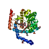
| ||||||||
|---|---|---|---|---|---|---|---|---|---|
| 1 | 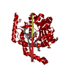
| ||||||||
| Unit cell |
|
-Components
-Protein / Sugars , 2 types, 3 molecules B
| #1: Protein | Mass: 38133.590 Da / Num. of mol.: 1 / Mutation: E127A Source method: isolated from a genetically manipulated source Source: (gene. exp.) Acinetobacter baumannii NIPH 410 (bacteria)Gene: F910_00940 / Production host: |
|---|---|
| #4: Sugar |  Type: D-saccharide / Mass: 510.615 Da / Num. of mol.: 2 / Source method: obtained synthetically / Formula: C24H46O11 / Comment: detergent*YM Type: D-saccharide / Mass: 510.615 Da / Num. of mol.: 2 / Source method: obtained synthetically / Formula: C24H46O11 / Comment: detergent*YM |
-Non-polymers , 4 types, 113 molecules 


| #2: Chemical | ChemComp-SO4 /  Mass: 96.063 Da / Num. of mol.: 4 / Source method: obtained synthetically / Formula: SO4 Mass: 96.063 Da / Num. of mol.: 4 / Source method: obtained synthetically / Formula: SO4#3: Chemical |  Mass: 92.094 Da / Num. of mol.: 3 / Source method: obtained synthetically / Formula: C3H8O3 Mass: 92.094 Da / Num. of mol.: 3 / Source method: obtained synthetically / Formula: C3H8O3#5: Chemical | ChemComp-11A / |  Mass: 186.291 Da / Num. of mol.: 1 / Source method: obtained synthetically / Formula: C11H22O2 / Comment: antifungal*YM Mass: 186.291 Da / Num. of mol.: 1 / Source method: obtained synthetically / Formula: C11H22O2 / Comment: antifungal*YM#6: Water | ChemComp-HOH / | Mass: 18.015 Da / Num. of mol.: 105 / Source method: isolated from a natural source / Formula: H2O |
|---|
-Experimental details
-Experiment
| Experiment | Method: X-RAY DIFFRACTION / Number of used crystals: 1 |
|---|
- Sample preparation
| Crystal | Density Matthews: 2.56 Å3/Da / Density % sol: 51.94 % / Description: Small guitar pick shaped crystals. |
|---|---|
| Crystal grow | Temperature: 293 K / Method: vapor diffusion, sitting drop / pH: 7.5 / Details: 200 mM KBr 2.2 M (NH4)2SO4 |
-Data collection
| Diffraction | Mean temperature: 80 K |
|---|---|
| Diffraction source | Source: SYNCHROTRON / Site: ALS  / Beamline: 5.0.2 / Wavelength: 1 Å / Beamline: 5.0.2 / Wavelength: 1 Å |
| Detector | Type: ADSC QUANTUM 315r / Detector: CCD / Date: Apr 8, 2016 |
| Radiation | Protocol: SINGLE WAVELENGTH / Monochromatic (M) / Laue (L): M / Scattering type: x-ray |
| Radiation wavelength | Wavelength: 1 Å / Relative weight: 1 |
| Reflection | Resolution: 1.9→72.74 Å / Num. obs: 30262 / % possible obs: 100 % / Redundancy: 1.9 % / CC1/2: 0.998 / Net I/σ(I): 7.95 |
- Processing
| Software |
| ||||||||||||||||||||||||||||||||||||||||||
|---|---|---|---|---|---|---|---|---|---|---|---|---|---|---|---|---|---|---|---|---|---|---|---|---|---|---|---|---|---|---|---|---|---|---|---|---|---|---|---|---|---|---|---|
| Refinement | Method to determine structure: MOLECULAR REPLACEMENT Starting model: 5KN7 Resolution: 1.9→72.735 Å / SU ML: 0.3 / Cross valid method: FREE R-VALUE / σ(F): 1.33 / Phase error: 29.79
| ||||||||||||||||||||||||||||||||||||||||||
| Solvent computation | Shrinkage radii: 0.9 Å / VDW probe radii: 1.11 Å | ||||||||||||||||||||||||||||||||||||||||||
| Displacement parameters | Biso max: 104.9 Å2 / Biso mean: 32.284 Å2 / Biso min: 10.68 Å2 | ||||||||||||||||||||||||||||||||||||||||||
| Refinement step | Cycle: final / Resolution: 1.9→72.735 Å
| ||||||||||||||||||||||||||||||||||||||||||
| Refine LS restraints |
| ||||||||||||||||||||||||||||||||||||||||||
| LS refinement shell | Refine-ID: X-RAY DIFFRACTION / Total num. of bins used: 6 / % reflection obs: 100 %
|
