[English] 日本語
 Yorodumi
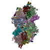
- PDB-5kai: NH3-bound RT XFEL structure of Photosystem II 500 ms after the 2n... -
+ Open data
- Basic information
| Entry | Database: PDB / ID: 5kai | ||||||||||||||||||||||||||||||||||||||||||||||||||||||||||||||||||
|---|---|---|---|---|---|---|---|---|---|---|---|---|---|---|---|---|---|---|---|---|---|---|---|---|---|---|---|---|---|---|---|---|---|---|---|---|---|---|---|---|---|---|---|---|---|---|---|---|---|---|---|---|---|---|---|---|---|---|---|---|---|---|---|---|---|---|---|
| Title | NH3-bound RT XFEL structure of Photosystem II 500 ms after the 2nd illumination (2F) at 2.8 A resolution | ||||||||||||||||||||||||||||||||||||||||||||||||||||||||||||||||||
 Components Components |
| ||||||||||||||||||||||||||||||||||||||||||||||||||||||||||||||||||
 Keywords Keywords | ELECTRON TRANSPORT / PHOTOSYSTEMS / TRANSMEMBRANE / ROOM TEMPERATURE | ||||||||||||||||||||||||||||||||||||||||||||||||||||||||||||||||||
| Function / homology |  Function and homology information Function and homology informationphotosystem II oxygen evolving complex / photosystem II assembly / oxygen evolving activity / photosystem II stabilization / photosystem II reaction center / photosystem II / oxidoreductase activity, acting on diphenols and related substances as donors, oxygen as acceptor / photosynthetic electron transport chain / : / photosystem II ...photosystem II oxygen evolving complex / photosystem II assembly / oxygen evolving activity / photosystem II stabilization / photosystem II reaction center / photosystem II / oxidoreductase activity, acting on diphenols and related substances as donors, oxygen as acceptor / photosynthetic electron transport chain / : / photosystem II / response to herbicide / extrinsic component of membrane / plasma membrane-derived thylakoid membrane / chlorophyll binding / photosynthetic electron transport in photosystem II / phosphate ion binding / photosynthesis, light reaction / photosynthesis / respiratory electron transport chain / manganese ion binding / electron transfer activity / protein stabilization / iron ion binding / heme binding / metal ion binding Similarity search - Function | ||||||||||||||||||||||||||||||||||||||||||||||||||||||||||||||||||
| Biological species |   Thermosynechococcus elongatus (bacteria) Thermosynechococcus elongatus (bacteria) | ||||||||||||||||||||||||||||||||||||||||||||||||||||||||||||||||||
| Method |  X-RAY DIFFRACTION / X-RAY DIFFRACTION /  FREE ELECTRON LASER / FREE ELECTRON LASER /  MOLECULAR REPLACEMENT / Resolution: 2.80000914773 Å MOLECULAR REPLACEMENT / Resolution: 2.80000914773 Å | ||||||||||||||||||||||||||||||||||||||||||||||||||||||||||||||||||
 Authors Authors | Young, I.D. / Ibrahim, M. / Chatterjee, R. / Gul, S. / Koroidov, S. / Brewster, A.S. / Tran, R. / Alonso-Mori, R. / Fuller, F. / Kroll, T. ...Young, I.D. / Ibrahim, M. / Chatterjee, R. / Gul, S. / Koroidov, S. / Brewster, A.S. / Tran, R. / Alonso-Mori, R. / Fuller, F. / Kroll, T. / Michels-Clark, T. / Laksmono, H. / Sierra, R.G. / Stan, C.A. / Saracini, C. / Bean, M.A. / Seuffert, I. / Sokaras, D. / Weng, T.-C. / Hunter, M.S. / Aquila, A. / Koglin, J.E. / Robinson, J. / Liang, M. / Boutet, S. / Lyubimov, A.Y. / Uervirojnangkoorn, M. / Moriarty, N.W. / Liebschner, D. / Afonine, P.V. / Waterman, D.G. / Evans, G. / Dobbek, H. / Weis, W.I. / Brunger, A.T. / Zwart, P.H. / Adams, P.D. / Zouni, A. / Messinger, J. / Bergmann, U. / Sauter, N.K. / Kern, J. / Yachandra, V.K. / Yano, J. | ||||||||||||||||||||||||||||||||||||||||||||||||||||||||||||||||||
| Funding support |  United States, United States,  France, France,  Germany, Germany,  Sweden, Sweden,  United Kingdom, 21items United Kingdom, 21items
| ||||||||||||||||||||||||||||||||||||||||||||||||||||||||||||||||||
 Citation Citation |  Journal: Nature / Year: 2016 Journal: Nature / Year: 2016Title: Structure of photosystem II and substrate binding at room temperature. Authors: Young, I.D. / Ibrahim, M. / Chatterjee, R. / Gul, S. / Fuller, F.D. / Koroidov, S. / Brewster, A.S. / Tran, R. / Alonso-Mori, R. / Kroll, T. / Michels-Clark, T. / Laksmono, H. / Sierra, R.G. ...Authors: Young, I.D. / Ibrahim, M. / Chatterjee, R. / Gul, S. / Fuller, F.D. / Koroidov, S. / Brewster, A.S. / Tran, R. / Alonso-Mori, R. / Kroll, T. / Michels-Clark, T. / Laksmono, H. / Sierra, R.G. / Stan, C.A. / Hussein, R. / Zhang, M. / Douthit, L. / Kubin, M. / de Lichtenberg, C. / Vo Pham, L. / Nilsson, H. / Cheah, M.H. / Shevela, D. / Saracini, C. / Bean, M.A. / Seuffert, I. / Sokaras, D. / Weng, T.C. / Pastor, E. / Weninger, C. / Fransson, T. / Lassalle, L. / Brauer, P. / Aller, P. / Docker, P.T. / Andi, B. / Orville, A.M. / Glownia, J.M. / Nelson, S. / Sikorski, M. / Zhu, D. / Hunter, M.S. / Lane, T.J. / Aquila, A. / Koglin, J.E. / Robinson, J. / Liang, M. / Boutet, S. / Lyubimov, A.Y. / Uervirojnangkoorn, M. / Moriarty, N.W. / Liebschner, D. / Afonine, P.V. / Waterman, D.G. / Evans, G. / Wernet, P. / Dobbek, H. / Weis, W.I. / Brunger, A.T. / Zwart, P.H. / Adams, P.D. / Zouni, A. / Messinger, J. / Bergmann, U. / Sauter, N.K. / Kern, J. / Yachandra, V.K. / Yano, J. #1: Journal: Acta Crystallogr.,Sect.D / Year: 2012 Title: Towards automated crystallographic structure refinement with phenix.refine. Authors: Afonine, P.V. / Grosse-Kunstleve, R.W. / Echols, N. / Headd, J.J. / Moriarty, N.W. / Mustyakimov, M. / Terwilliger, T.C. / Urzhumtsev, A. / Zwart, P.H. / Adams, P.D. #2: Journal: Acta Crystallogr D Biol Crystallogr / Year: 2010 Title: PHENIX: a comprehensive Python-based system for macromolecular structure solution. Authors: Paul D Adams / Pavel V Afonine / Gábor Bunkóczi / Vincent B Chen / Ian W Davis / Nathaniel Echols / Jeffrey J Headd / Li-Wei Hung / Gary J Kapral / Ralf W Grosse-Kunstleve / Airlie J McCoy ...Authors: Paul D Adams / Pavel V Afonine / Gábor Bunkóczi / Vincent B Chen / Ian W Davis / Nathaniel Echols / Jeffrey J Headd / Li-Wei Hung / Gary J Kapral / Ralf W Grosse-Kunstleve / Airlie J McCoy / Nigel W Moriarty / Robert Oeffner / Randy J Read / David C Richardson / Jane S Richardson / Thomas C Terwilliger / Peter H Zwart /  Abstract: Macromolecular X-ray crystallography is routinely applied to understand biological processes at a molecular level. However, significant time and effort are still required to solve and complete many ...Macromolecular X-ray crystallography is routinely applied to understand biological processes at a molecular level. However, significant time and effort are still required to solve and complete many of these structures because of the need for manual interpretation of complex numerical data using many software packages and the repeated use of interactive three-dimensional graphics. PHENIX has been developed to provide a comprehensive system for macromolecular crystallographic structure solution with an emphasis on the automation of all procedures. This has relied on the development of algorithms that minimize or eliminate subjective input, the development of algorithms that automate procedures that are traditionally performed by hand and, finally, the development of a framework that allows a tight integration between the algorithms. | ||||||||||||||||||||||||||||||||||||||||||||||||||||||||||||||||||
| History |
|
- Structure visualization
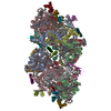
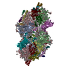
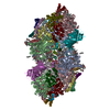
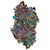
| Structure viewer | Molecule:  Molmil Molmil Jmol/JSmol Jmol/JSmol |
|---|
- Downloads & links
- Download
| PDBx/mmCIF format |  5kai.cif.gz 5kai.cif.gz | 1.3 MB | Display |  PDBx/mmCIF format PDBx/mmCIF format |
|---|---|---|---|---|
| PDB format |  pdb5kai.ent.gz pdb5kai.ent.gz | 1 MB | Display |  PDB format PDB format |
| PDBx/mmJSON format |  5kai.json.gz 5kai.json.gz | Tree view |  PDBx/mmJSON format PDBx/mmJSON format | |
| Others |  Other downloads Other downloads |
-Validation report
| Arichive directory |  https://data.pdbj.org/pub/pdb/validation_reports/ka/5kai https://data.pdbj.org/pub/pdb/validation_reports/ka/5kai ftp://data.pdbj.org/pub/pdb/validation_reports/ka/5kai ftp://data.pdbj.org/pub/pdb/validation_reports/ka/5kai | HTTPS FTP |
|---|
-Related structure data
| Related structure data | 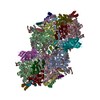 5kafC 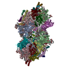 5tisC 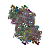 4pj0S 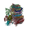 4ub6S C: citing same article ( S: Starting model for refinement |
|---|---|
| Similar structure data |
- Links
Links
- Assembly
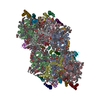
| Deposited unit | 
| ||||||||||||
|---|---|---|---|---|---|---|---|---|---|---|---|---|---|
| 1 |
| ||||||||||||
| Unit cell |
|
- Components
-Photosystem II ... , 17 types, 34 molecules AaBbCcDdHhIiJjKkLlMmOoTtUuYyXx...
| #1: Protein | Mass: 38265.625 Da / Num. of mol.: 2 / Fragment: UNP residues 1-344 / Source method: isolated from a natural source Source: (natural)   Thermosynechococcus elongatus (strain BP-1) (bacteria) Thermosynechococcus elongatus (strain BP-1) (bacteria)Strain: BP-1 / References: UniProt: P0A444, photosystem II #2: Protein | Mass: 56656.457 Da / Num. of mol.: 2 / Source method: isolated from a natural source Source: (natural)   Thermosynechococcus elongatus (strain BP-1) (bacteria) Thermosynechococcus elongatus (strain BP-1) (bacteria)Strain: BP-1 / References: UniProt: Q8DIQ1 #3: Protein | Mass: 50287.500 Da / Num. of mol.: 2 / Source method: isolated from a natural source Source: (natural)   Thermosynechococcus elongatus (strain BP-1) (bacteria) Thermosynechococcus elongatus (strain BP-1) (bacteria)Strain: BP-1 / References: UniProt: Q8DIF8 #4: Protein | Mass: 39388.156 Da / Num. of mol.: 2 / Source method: isolated from a natural source Source: (natural)   Thermosynechococcus elongatus (strain BP-1) (bacteria) Thermosynechococcus elongatus (strain BP-1) (bacteria)Strain: BP-1 / References: UniProt: Q8CM25, photosystem II #7: Protein | Mass: 7057.349 Da / Num. of mol.: 2 / Source method: isolated from a natural source Source: (natural)   Thermosynechococcus elongatus (strain BP-1) (bacteria) Thermosynechococcus elongatus (strain BP-1) (bacteria)Strain: BP-1 / References: UniProt: Q8DJ43 #8: Protein/peptide | Mass: 4438.255 Da / Num. of mol.: 2 / Source method: isolated from a natural source Source: (natural)   Thermosynechococcus elongatus (strain BP-1) (bacteria) Thermosynechococcus elongatus (strain BP-1) (bacteria)Strain: BP-1 / References: UniProt: Q8DJZ6 #9: Protein/peptide | Mass: 4105.908 Da / Num. of mol.: 2 / Source method: isolated from a natural source Source: (natural)   Thermosynechococcus elongatus (strain BP-1) (bacteria) Thermosynechococcus elongatus (strain BP-1) (bacteria)Strain: BP-1 / References: UniProt: P59087 #10: Protein/peptide | Mass: 5028.083 Da / Num. of mol.: 2 / Source method: isolated from a natural source Source: (natural)   Thermosynechococcus elongatus (strain BP-1) (bacteria) Thermosynechococcus elongatus (strain BP-1) (bacteria)Strain: BP-1 / References: UniProt: Q9F1K9 #11: Protein/peptide | Mass: 4299.044 Da / Num. of mol.: 2 / Source method: isolated from a natural source Source: (natural)   Thermosynechococcus elongatus (strain BP-1) (bacteria) Thermosynechococcus elongatus (strain BP-1) (bacteria)Strain: BP-1 / References: UniProt: Q8DIN8 #12: Protein/peptide | Mass: 4009.682 Da / Num. of mol.: 2 / Source method: isolated from a natural source Source: (natural)   Thermosynechococcus elongatus (strain BP-1) (bacteria) Thermosynechococcus elongatus (strain BP-1) (bacteria)Strain: BP-1 / References: UniProt: Q8DHA7 #13: Protein | Mass: 29637.443 Da / Num. of mol.: 2 / Source method: isolated from a natural source Source: (natural)   Thermosynechococcus elongatus (strain BP-1) (bacteria) Thermosynechococcus elongatus (strain BP-1) (bacteria)Strain: BP-1 / References: UniProt: P0A431 #14: Protein/peptide | Mass: 3906.738 Da / Num. of mol.: 2 / Source method: isolated from a natural source Source: (natural)   Thermosynechococcus elongatus (strain BP-1) (bacteria) Thermosynechococcus elongatus (strain BP-1) (bacteria)Strain: BP-1 / References: UniProt: Q8DIQ0 #15: Protein | Mass: 15030.986 Da / Num. of mol.: 2 / Source method: isolated from a natural source Source: (natural)   Thermosynechococcus elongatus (strain BP-1) (bacteria) Thermosynechococcus elongatus (strain BP-1) (bacteria)Strain: BP-1 / References: UniProt: Q9F1L5 #17: Protein/peptide | Mass: 5039.143 Da / Num. of mol.: 2 / Source method: isolated from a natural source Source: (natural)   Thermosynechococcus elongatus (strain BP-1) (bacteria) Thermosynechococcus elongatus (strain BP-1) (bacteria)Strain: BP-1 / References: UniProt: Q8DJI1 #18: Protein/peptide | Mass: 4322.226 Da / Num. of mol.: 2 / Source method: isolated from a natural source Source: (natural)   Thermosynechococcus elongatus (strain BP-1) (bacteria) Thermosynechococcus elongatus (strain BP-1) (bacteria)Strain: BP-1 / References: UniProt: Q9F1R6 #19: Protein | Mass: 6766.187 Da / Num. of mol.: 2 / Source method: isolated from a natural source Source: (natural)   Thermosynechococcus elongatus (strain BP-1) (bacteria) Thermosynechococcus elongatus (strain BP-1) (bacteria)Strain: BP-1 / References: UniProt: Q8DHJ2 #20: Protein/peptide | Mass: 4590.648 Da / Num. of mol.: 2 / Source method: isolated from a natural source Source: (natural)   Thermosynechococcus elongatus (strain BP-1) (bacteria) Thermosynechococcus elongatus (strain BP-1) (bacteria)Strain: BP-1 / References: UniProt: Q8DKM3 |
|---|
-Cytochrome b559 subunit ... , 2 types, 4 molecules EeFf
| #5: Protein | Mass: 9580.840 Da / Num. of mol.: 2 / Source method: isolated from a natural source Source: (natural)   Thermosynechococcus elongatus (strain BP-1) (bacteria) Thermosynechococcus elongatus (strain BP-1) (bacteria)Strain: BP-1 / References: UniProt: Q8DIP0 #6: Protein/peptide | Mass: 5067.900 Da / Num. of mol.: 2 / Source method: isolated from a natural source Source: (natural)   Thermosynechococcus elongatus (strain BP-1) (bacteria) Thermosynechococcus elongatus (strain BP-1) (bacteria)Strain: BP-1 / References: UniProt: Q8DIN9 |
|---|
-Protein / Sugars , 2 types, 10 molecules Vv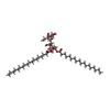

| #16: Protein | Mass: 18046.943 Da / Num. of mol.: 2 / Source method: isolated from a natural source Source: (natural)   Thermosynechococcus elongatus (strain BP-1) (bacteria) Thermosynechococcus elongatus (strain BP-1) (bacteria)Strain: BP-1 / References: UniProt: P0A386 #33: Sugar | ChemComp-DGD / |
|---|
-Non-polymers , 15 types, 279 molecules 



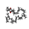
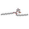
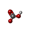


| #21: Chemical | | #22: Chemical | #23: Chemical | ChemComp-LMG / #24: Chemical | ChemComp-CL / #25: Chemical | ChemComp-CLA / #26: Chemical | ChemComp-PHO / #27: Chemical | ChemComp-BCR / #28: Chemical | ChemComp-PL9 / #29: Chemical | ChemComp-SQD / #30: Chemical | ChemComp-UNL / Mass: 616.487 Da / Num. of mol.: 23 / Source method: obtained synthetically #31: Chemical | #32: Chemical | ChemComp-LHG / #34: Chemical | #35: Chemical | #36: Water | ChemComp-HOH / | |
|---|
-Details
| Has protein modification | Y |
|---|
-Experimental details
-Experiment
| Experiment | Method:  X-RAY DIFFRACTION / Number of used crystals: 1 X-RAY DIFFRACTION / Number of used crystals: 1 |
|---|
- Sample preparation
| Crystal | Density Matthews: 3.71 Å3/Da / Density % sol: 66.86 % |
|---|---|
| Crystal grow | Temperature: 293 K / Method: batch mode / pH: 7.5 Details: Crystal growth was as described in Kern et al. 2005, Biochim. Biophys. Acta-Bioenergetics and Ibrahim et al. 2015, Struct. Dyn. 2 (4), 041705, substituting C12E8 for beta-DM and Betaine for glycerol |
-Data collection
| Diffraction | Mean temperature: 293 K | |||||||||||||||||||||||||||||||||||||||||||||||||||||||||||||||||||||||||||||||||||||||||||||||||||||||||
|---|---|---|---|---|---|---|---|---|---|---|---|---|---|---|---|---|---|---|---|---|---|---|---|---|---|---|---|---|---|---|---|---|---|---|---|---|---|---|---|---|---|---|---|---|---|---|---|---|---|---|---|---|---|---|---|---|---|---|---|---|---|---|---|---|---|---|---|---|---|---|---|---|---|---|---|---|---|---|---|---|---|---|---|---|---|---|---|---|---|---|---|---|---|---|---|---|---|---|---|---|---|---|---|---|---|---|
| Diffraction source | Source:  FREE ELECTRON LASER / Site: FREE ELECTRON LASER / Site:  SLAC LCLS SLAC LCLS  / Beamline: CXI / Wavelength: 1.7492 Å / Beamline: CXI / Wavelength: 1.7492 Å | |||||||||||||||||||||||||||||||||||||||||||||||||||||||||||||||||||||||||||||||||||||||||||||||||||||||||
| Detector | Type: CS-PAD CXI-1 / Detector: PIXEL / Date: Nov 28, 2014 / Details: 1 micron KB mirror | |||||||||||||||||||||||||||||||||||||||||||||||||||||||||||||||||||||||||||||||||||||||||||||||||||||||||
| Radiation | Protocol: SINGLE WAVELENGTH / Monochromatic (M) / Laue (L): M / Scattering type: x-ray | |||||||||||||||||||||||||||||||||||||||||||||||||||||||||||||||||||||||||||||||||||||||||||||||||||||||||
| Radiation wavelength | Wavelength: 1.7492 Å / Relative weight: 1 | |||||||||||||||||||||||||||||||||||||||||||||||||||||||||||||||||||||||||||||||||||||||||||||||||||||||||
| Reflection | Resolution: 2.8→43.569812 Å / Num. obs: 213400 / % possible obs: 99 % / Redundancy: 13.7 % / Biso Wilson estimate: 60.4069710515 Å2 / CC1/2: 0.524 / Net I/σ(I): 11.62 | |||||||||||||||||||||||||||||||||||||||||||||||||||||||||||||||||||||||||||||||||||||||||||||||||||||||||
| Reflection shell |
|
- Processing
| Software |
| ||||||||||||||||||||||||||||||||||||||||||||||||||||||||||||||||||||||||||||||||||||||||||||||||||
|---|---|---|---|---|---|---|---|---|---|---|---|---|---|---|---|---|---|---|---|---|---|---|---|---|---|---|---|---|---|---|---|---|---|---|---|---|---|---|---|---|---|---|---|---|---|---|---|---|---|---|---|---|---|---|---|---|---|---|---|---|---|---|---|---|---|---|---|---|---|---|---|---|---|---|---|---|---|---|---|---|---|---|---|---|---|---|---|---|---|---|---|---|---|---|---|---|---|---|---|
| Refinement | Method to determine structure:  MOLECULAR REPLACEMENT MOLECULAR REPLACEMENTStarting model: PSII monomer composed of 4UB6 monomer and additional chain from 4PJ0 Resolution: 2.80000914773→43.5698116774 Å / SU ML: 0.504121620111 / Cross valid method: FREE R-VALUE / σ(F): 1.32517238381 / Phase error: 34.5352448831 Stereochemistry target values: GeoStd + Monomer Library + CDL v1.2
| ||||||||||||||||||||||||||||||||||||||||||||||||||||||||||||||||||||||||||||||||||||||||||||||||||
| Solvent computation | Shrinkage radii: 0.9 Å / VDW probe radii: 1.11 Å / Solvent model: FLAT BULK SOLVENT MODEL | ||||||||||||||||||||||||||||||||||||||||||||||||||||||||||||||||||||||||||||||||||||||||||||||||||
| Displacement parameters | Biso mean: 56.5229924032 Å2 | ||||||||||||||||||||||||||||||||||||||||||||||||||||||||||||||||||||||||||||||||||||||||||||||||||
| Refinement step | Cycle: LAST / Resolution: 2.80000914773→43.5698116774 Å
| ||||||||||||||||||||||||||||||||||||||||||||||||||||||||||||||||||||||||||||||||||||||||||||||||||
| Refine LS restraints |
| ||||||||||||||||||||||||||||||||||||||||||||||||||||||||||||||||||||||||||||||||||||||||||||||||||
| LS refinement shell |
|
 Movie
Movie Controller






 PDBj
PDBj


