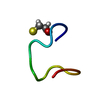
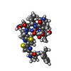
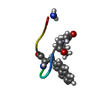
The asymmetric unit is rA2, however the input RNA polymer is rA7, and this forms a continual parallel poly(rA) double helix structure. This structure is referred to as a "staggered zipper" since the rA7 strands overlap in a many different registers relative to their opposite, paired strands, to form the helix.
-
Components
#1: RNA chain
RNA7-mer
Mass: 2259.483 Da / Num. of mol.: 1 / Source method: obtained synthetically / Source: (synth.) synthetic construct (others)
Mass: 18.015 Da / Num. of mol.: 8 / Source method: isolated from a natural source / Formula: H2O
-
Experimental details
-
Experiment
Experiment
Method: X-RAY DIFFRACTION / Number of used crystals: 1
-
Sample preparation
Crystal
Density Matthews: 1.33 Å3/Da / Density % sol: 8 %
Crystal grow
Temperature: 293.15 K / Method: vapor diffusion, hanging drop / pH: 3.5 Details: rA7 dissolved in water to 10 mM and was diluted 1/10 v/v in 0.75% PEG 1000. One uL of this was mixed with 1 uL of the following reservoir solution: (0.24 M ammonium sulfate and 0.06 M citric ...Details: rA7 dissolved in water to 10 mM and was diluted 1/10 v/v in 0.75% PEG 1000. One uL of this was mixed with 1 uL of the following reservoir solution: (0.24 M ammonium sulfate and 0.06 M citric acid pH 3.5) to generate a hanging drop over 1 mL of this reservoir solution.
In the structure databanks used in Yorodumi, some data are registered as the other names, "COVID-19 virus" and "2019-nCoV". Here are the details of the virus and the list of structure data.
Jan 31, 2019. EMDB accession codes are about to change! (news from PDBe EMDB page)
EMDB accession codes are about to change! (news from PDBe EMDB page)
The allocation of 4 digits for EMDB accession codes will soon come to an end. Whilst these codes will remain in use, new EMDB accession codes will include an additional digit and will expand incrementally as the available range of codes is exhausted. The current 4-digit format prefixed with “EMD-” (i.e. EMD-XXXX) will advance to a 5-digit format (i.e. EMD-XXXXX), and so on. It is currently estimated that the 4-digit codes will be depleted around Spring 2019, at which point the 5-digit format will come into force.
The EM Navigator/Yorodumi systems omit the EMD- prefix.
Related info.:Q: What is EMD? / ID/Accession-code notation in Yorodumi/EM Navigator
Yorodumi is a browser for structure data from EMDB, PDB, SASBDB, etc.
This page is also the successor to EM Navigator detail page, and also detail information page/front-end page for Omokage search.
The word "yorodu" (or yorozu) is an old Japanese word meaning "ten thousand". "mi" (miru) is to see.
Related info.:EMDB / PDB / SASBDB / Comparison of 3 databanks / Yorodumi Search / Aug 31, 2016. New EM Navigator & Yorodumi / Yorodumi Papers / Jmol/JSmol / Function and homology information / Changes in new EM Navigator and Yorodumi
 Movie
Movie Controller

 Yorodumi
 Open data
 Basic information
 Components
 Keywords
 Function and homology information
 X-RAY DIFFRACTION /
  Authors
 United States, 3items
  Citation
 Structure visualization
 Downloads & links
 Other downloads
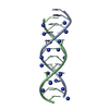
 PDBj
PDBj
 Assembly


 Mass: 18.038 Da / Num. of mol.: 1 / Source method: obtained synthetically / Formula: H4N
 Mass: 18.015 Da / Num. of mol.: 8 / Source method: isolated from a natural source / Formula: H2O
 Sample preparation
 Processing