 Movie
Movie Controller

[English] 日本語
 Yorodumi
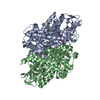
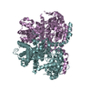
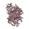
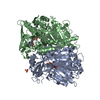
- PDB-5k3i: Crystal structure of Acyl-CoA oxidase-1 in Caenorhabditis elegans... -
+ Open data

- Basic information
| Entry | Database: PDB / ID: 5k3i | ||||||||||||||||||
|---|---|---|---|---|---|---|---|---|---|---|---|---|---|---|---|---|---|---|---|
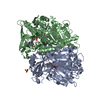
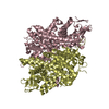
| Title | Crystal structure of Acyl-CoA oxidase-1 in Caenorhabditis elegans complexed with FAD and ATP | ||||||||||||||||||
 Components Components | Acyl-coenzyme A oxidase | ||||||||||||||||||
 Keywords Keywords | OXIDOREDUCTASE / dauer pheromone / ascarosides / b-oxidation / ATP | ||||||||||||||||||
| Function / homology |  Function and homology information Function and homology informationascaroside biosynthetic process / Beta-oxidation of pristanoyl-CoA / Synthesis of bile acids and bile salts via 7alpha-hydroxycholesterol / Oxidoreductases; Acting on the CH-CH group of donors; With oxygen as acceptor / acyl-CoA oxidase / acyl-CoA oxidase activity / pheromone biosynthetic process / fatty acid beta-oxidation using acyl-CoA oxidase / peroxisomal matrix / FAD binding ...ascaroside biosynthetic process / Beta-oxidation of pristanoyl-CoA / Synthesis of bile acids and bile salts via 7alpha-hydroxycholesterol / Oxidoreductases; Acting on the CH-CH group of donors; With oxygen as acceptor / acyl-CoA oxidase / acyl-CoA oxidase activity / pheromone biosynthetic process / fatty acid beta-oxidation using acyl-CoA oxidase / peroxisomal matrix / FAD binding / fatty acid binding / flavin adenine dinucleotide binding / peroxisome / ATP binding Similarity search - Function | ||||||||||||||||||
| Biological species |  | ||||||||||||||||||
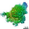
| Method |  X-RAY DIFFRACTION / SYNCHROTRON / MOLECULAR REPLACEMENT / Resolution: 2.683 Å X-RAY DIFFRACTION / SYNCHROTRON / MOLECULAR REPLACEMENT / Resolution: 2.683 Å | ||||||||||||||||||
 Authors Authors | Zhang, X. / Li, K. / Jones, R.A. / Bruner, S.D. / Butcher, R.A. | ||||||||||||||||||
| Funding support |  United States, 5items United States, 5items
| ||||||||||||||||||
 Citation Citation | Journal: Proc.Natl.Acad.Sci.USA / Year: 2016 Title: Structural characterization of acyl-CoA oxidases reveals a direct link between pheromone biosynthesis and metabolic state in Caenorhabditis elegans. Authors: Zhang, X. / Li, K. / Jones, R.A. / Bruner, S.D. / Butcher, R.A. | ||||||||||||||||||
| History |
|
- Structure visualization
| Structure viewer | Molecule: MolmilJmol/JSmol |
|---|
- Downloads & links
-Download
| PDBx/mmCIF format | 5k3i.cif.gz | 1 MB | Display | PDBx/mmCIF format |
|---|---|---|---|---|
| PDB format | pdb5k3i.ent.gz | 847.8 KB | Display | PDB format |
| PDBx/mmJSON format | 5k3i.json.gz | Tree view | PDBx/mmJSON format | |
| Others |  Other downloads Other downloads |
-Validation report
| Arichive directory | https://data.pdbj.org/pub/pdb/validation_reports/k3/5k3iftp://data.pdbj.org/pub/pdb/validation_reports/k3/5k3i | HTTPS FTP |
|---|
-Related structure data
| Related structure data |  5k3gSC  5k3hC  5k3jC S: Starting model for refinement C: citing same article ( |
|---|---|
| Similar structure data |


-Links
 PDBj
PDBj- Assembly
| Deposited unit | 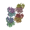
| ||||||||
|---|---|---|---|---|---|---|---|---|---|
| 1 | 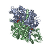
| ||||||||
| 2 | 
| ||||||||
| 3 | 
| ||||||||
| 4 | 
| ||||||||
| Unit cell |
|
-Components
| #1: Protein | Mass: 77499.820 Da / Num. of mol.: 8 / Mutation: E434A Source method: isolated from a genetically manipulated source Source: (gene. exp.)  #2: Chemical | ChemComp-FAD /   Mass: 785.550 Da / Num. of mol.: 8 / Source method: obtained synthetically / Formula: C27H33N9O15P2 / Comment: FAD*YM Mass: 785.550 Da / Num. of mol.: 8 / Source method: obtained synthetically / Formula: C27H33N9O15P2 / Comment: FAD*YM#3: Chemical | ChemComp-ATP /   Mass: 507.181 Da / Num. of mol.: 8 / Source method: obtained synthetically / Formula: C10H16N5O13P3 / Comment: ATP, energy-carrying molecule*YM Mass: 507.181 Da / Num. of mol.: 8 / Source method: obtained synthetically / Formula: C10H16N5O13P3 / Comment: ATP, energy-carrying molecule*YM#4: Chemical | ChemComp-MG /   Mass: 24.305 Da / Num. of mol.: 8 / Source method: obtained synthetically / Formula: Mg Mass: 24.305 Da / Num. of mol.: 8 / Source method: obtained synthetically / Formula: Mg#5: Water | ChemComp-HOH / |  Mass: 18.015 Da / Num. of mol.: 601 / Source method: isolated from a natural source / Formula: H2O Mass: 18.015 Da / Num. of mol.: 601 / Source method: isolated from a natural source / Formula: H2O |
|---|
-Experimental details
-Experiment
| Experiment | Method: X-RAY DIFFRACTION / Number of used crystals: 1 |
|---|
- Sample preparation
| Crystal | Density Matthews: 2.43 Å3/Da / Density % sol: 49.4 % |
|---|---|
| Crystal grow | Temperature: 298 K / Method: vapor diffusion, hanging drop Details: 0.4 M NaCl, 0.1 M HEPES pH 7.4, 18% w/v PEG 8000 and 8% v/v glycerol PH range: 7.4 |
-Data collection
| Diffraction | Mean temperature: 100 K |
|---|---|
| Diffraction source | Source: SYNCHROTRON / Site: APS / Beamline: 21-ID-G / Wavelength: 0.9787 Å |
| Detector | Type: MARMOSAIC 300 mm CCD / Detector: CCD / Date: Mar 13, 2015 |
| Radiation | Monochromator: Diamond [111] / Protocol: SINGLE WAVELENGTH / Monochromatic (M) / Laue (L): M / Scattering type: x-ray |
| Radiation wavelength | Wavelength: 0.9787 Å / Relative weight: 1 |
| Reflection | Resolution: 2.63→38.736 Å / Num. obs: 163296 / % possible obs: 100 % / Redundancy: 3.8 % / Biso Wilson estimate: 32.3 Å2 / Rmerge(I) obs: 0.14 / Rsym value: 0.17 / Net I/σ(I): 8.4 |
| Reflection shell | Resolution: 2.63→2.78 Å / Redundancy: 3.8 % / Rmerge(I) obs: 0.61 / Mean I/σ(I) obs: 0.7 / % possible all: 98.3 |
- Processing
| Software |
| |||||||||||||||||||||||||||||||||||||||||||||||||||||||||||||||||||||||||||||||||||||||||||||||||||||||||||||||||||||||||||||||||||||||||||||||||||
|---|---|---|---|---|---|---|---|---|---|---|---|---|---|---|---|---|---|---|---|---|---|---|---|---|---|---|---|---|---|---|---|---|---|---|---|---|---|---|---|---|---|---|---|---|---|---|---|---|---|---|---|---|---|---|---|---|---|---|---|---|---|---|---|---|---|---|---|---|---|---|---|---|---|---|---|---|---|---|---|---|---|---|---|---|---|---|---|---|---|---|---|---|---|---|---|---|---|---|---|---|---|---|---|---|---|---|---|---|---|---|---|---|---|---|---|---|---|---|---|---|---|---|---|---|---|---|---|---|---|---|---|---|---|---|---|---|---|---|---|---|---|---|---|---|---|---|---|---|
| Refinement | Method to determine structure: MOLECULAR REPLACEMENT Starting model: 5K3G Resolution: 2.683→38.736 Å / Cross valid method: FREE R-VALUE / σ(F): 1.34 / Phase error: 28.56 / Stereochemistry target values: TWIN_LSQ_F
| |||||||||||||||||||||||||||||||||||||||||||||||||||||||||||||||||||||||||||||||||||||||||||||||||||||||||||||||||||||||||||||||||||||||||||||||||||
| Solvent computation | Shrinkage radii: 0.9 Å / VDW probe radii: 1.11 Å / Solvent model: FLAT BULK SOLVENT MODEL | |||||||||||||||||||||||||||||||||||||||||||||||||||||||||||||||||||||||||||||||||||||||||||||||||||||||||||||||||||||||||||||||||||||||||||||||||||
| Refinement step | Cycle: LAST / Resolution: 2.683→38.736 Å
| |||||||||||||||||||||||||||||||||||||||||||||||||||||||||||||||||||||||||||||||||||||||||||||||||||||||||||||||||||||||||||||||||||||||||||||||||||
| Refine LS restraints |
| |||||||||||||||||||||||||||||||||||||||||||||||||||||||||||||||||||||||||||||||||||||||||||||||||||||||||||||||||||||||||||||||||||||||||||||||||||
| LS refinement shell |
|
