| Entry | Database: PDB / ID: 5jth
|
|---|
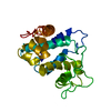
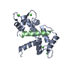
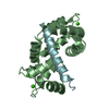
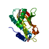
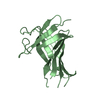
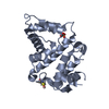
| Title | Crystal structure of E67A calmodulin - CaM:RM20 analog complex |
|---|
 Components Components | - Calmodulin
- Myosin light chain kinase, smooth muscle
|
|---|
 Keywords Keywords | TRANSFERASE / E67A calmodulin / calcium signal transduction / protein kinase / myosin light chain kinase |
|---|
| Function / homology |  Function and homology information Function and homology information
aorta smooth muscle tissue morphogenesis / tonic smooth muscle contraction / myosin-light-chain kinase / myosin light chain kinase activity / : / : / : / : / positive regulation of protein autophosphorylation / : ...aorta smooth muscle tissue morphogenesis / tonic smooth muscle contraction / myosin-light-chain kinase / myosin light chain kinase activity / : / : / : / : / positive regulation of protein autophosphorylation / : / cellular hypotonic response / negative regulation of peptidyl-threonine phosphorylation / bleb assembly / : / type 3 metabotropic glutamate receptor binding / positive regulation of calcium ion transport / positive regulation of peptidyl-threonine phosphorylation / positive regulation of DNA binding / CaM pathway / Cam-PDE 1 activation / Sodium/Calcium exchangers / Calmodulin induced events / Reduction of cytosolic Ca++ levels / Activation of Ca-permeable Kainate Receptor / CREB1 phosphorylation through the activation of CaMKII/CaMKK/CaMKIV cascasde / Loss of phosphorylation of MECP2 at T308 / CREB1 phosphorylation through the activation of Adenylate Cyclase / negative regulation of high voltage-gated calcium channel activity / PKA activation / CaMK IV-mediated phosphorylation of CREB / positive regulation of wound healing / Glycogen breakdown (glycogenolysis) / CLEC7A (Dectin-1) induces NFAT activation / negative regulation of ryanodine-sensitive calcium-release channel activity / response to corticosterone / organelle localization by membrane tethering / Activation of RAC1 downstream of NMDARs / : / autophagosome membrane docking / negative regulation of calcium ion export across plasma membrane / regulation of ryanodine-sensitive calcium-release channel activity / regulation of cardiac muscle cell action potential / regulation of synaptic vesicle exocytosis / presynaptic endocytosis / Synthesis of IP3 and IP4 in the cytosol / positive regulation of protein serine/threonine kinase activity / Phase 0 - rapid depolarisation / cleavage furrow / Negative regulation of NMDA receptor-mediated neuronal transmission / Unblocking of NMDA receptors, glutamate binding and activation / RHO GTPases activate PAKs / calcineurin-mediated signaling / nitric-oxide synthase binding / regulation of cell communication by electrical coupling involved in cardiac conduction / smooth muscle contraction / Ion transport by P-type ATPases / adenylate cyclase binding / Uptake and function of anthrax toxins / protein phosphatase activator activity / Long-term potentiation / Calcineurin activates NFAT / Regulation of MECP2 expression and activity / DARPP-32 events / Smooth Muscle Contraction / regulation of synaptic vesicle endocytosis / detection of calcium ion / regulation of cardiac muscle contraction / catalytic complex / RHO GTPases activate IQGAPs / phosphatidylinositol 3-kinase binding / positive regulation of nitric-oxide synthase activity / activation of adenylate cyclase activity / calcium channel inhibitor activity / presynaptic cytosol / Activation of AMPK downstream of NMDARs / enzyme regulator activity / cellular response to interferon-beta / regulation of release of sequestered calcium ion into cytosol by sarcoplasmic reticulum / Ion homeostasis / eNOS activation / stress fiber / Tetrahydrobiopterin (BH4) synthesis, recycling, salvage and regulation / Protein methylation / titin binding / regulation of cardiac muscle contraction by regulation of the release of sequestered calcium ion / regulation of calcium-mediated signaling / voltage-gated potassium channel complex / FCERI mediated Ca+2 mobilization / calcium channel complex / substantia nigra development / FCGR3A-mediated IL10 synthesis / regulation of heart rate / Antigen activates B Cell Receptor (BCR) leading to generation of second messengers / Ras activation upon Ca2+ influx through NMDA receptor / calyx of Held / nitric-oxide synthase regulator activity / adenylate cyclase activator activity / VEGFR2 mediated cell proliferation / VEGFR2 mediated vascular permeability / regulation of cytokinesisSimilarity search - Function Myosin Light Chain Kinase 1, Kinase domain / Unstructured linker between I-set domains 2 and 3 on MYLCK / Immunoglobulin I-set / Immunoglobulin I-set domain / : / Fibronectin type III domain / Fibronectin type 3 domain / Immunoglobulin subtype 2 / Immunoglobulin C-2 Type / Fibronectin type-III domain profile. ...Myosin Light Chain Kinase 1, Kinase domain / Unstructured linker between I-set domains 2 and 3 on MYLCK / Immunoglobulin I-set / Immunoglobulin I-set domain / : / Fibronectin type III domain / Fibronectin type 3 domain / Immunoglobulin subtype 2 / Immunoglobulin C-2 Type / Fibronectin type-III domain profile. / EF-hand domain pair / Fibronectin type III / Fibronectin type III superfamily / EF-hand, calcium binding motif / EF-Hand 1, calcium-binding site / EF-hand calcium-binding domain. / EF-hand calcium-binding domain profile. / EF-hand domain / EF-hand domain pair / Immunoglobulin subtype / Immunoglobulin / Ig-like domain profile. / Immunoglobulin-like domain / Immunoglobulin-like domain superfamily / Serine/threonine-protein kinase, active site / Serine/Threonine protein kinases active-site signature. / Protein kinase domain / Serine/Threonine protein kinases, catalytic domain / Protein kinase, ATP binding site / Protein kinases ATP-binding region signature. / Immunoglobulin-like fold / Protein kinase domain profile. / Protein kinase domain / Protein kinase-like domain superfamilySimilarity search - Domain/homology |
|---|
| Biological species |  Homo sapiens (human) Homo sapiens (human) |
|---|
| Method |  X-RAY DIFFRACTION / SYNCHROTRON / Resolution: 1.84 Å X-RAY DIFFRACTION / SYNCHROTRON / Resolution: 1.84 Å |
|---|
 Authors Authors | Grum-Tokars, V.L. / Minasov, G. / Anderson, W.F. / Watterson, D.M. |
|---|
 Citation Citation | Journal: To Be Published
Title: Crystal structure of E67A calmodulin - CaM:RM20 analog complex
Authors: Grum-Tokars, V.L. / Minasov, G. / Anderson, W.F. / Watterson, D.M. |
|---|
| History | | Deposition | May 9, 2016 | Deposition site: RCSB / Processing site: RCSB |
|---|
| Revision 1.0 | Jul 19, 2017 | Provider: repository / Type: Initial release |
|---|
| Revision 1.1 | Mar 6, 2024 | Group: Data collection / Database references ...Data collection / Database references / Derived calculations / Structure summary
Category: chem_comp_atom / chem_comp_bond ...chem_comp_atom / chem_comp_bond / citation / database_2 / struct / struct_conn / struct_conn_type
Item: _citation.title / _database_2.pdbx_DOI ..._citation.title / _database_2.pdbx_DOI / _database_2.pdbx_database_accession / _struct.title / _struct_conn.conn_type_id / _struct_conn.id / _struct_conn.pdbx_dist_value / _struct_conn.pdbx_leaving_atom_flag / _struct_conn.pdbx_ptnr1_label_alt_id / _struct_conn.ptnr1_auth_asym_id / _struct_conn.ptnr1_auth_comp_id / _struct_conn.ptnr1_auth_seq_id / _struct_conn.ptnr1_label_asym_id / _struct_conn.ptnr1_label_atom_id / _struct_conn.ptnr1_label_comp_id / _struct_conn.ptnr1_label_seq_id / _struct_conn.ptnr2_auth_asym_id / _struct_conn.ptnr2_auth_comp_id / _struct_conn.ptnr2_auth_seq_id / _struct_conn.ptnr2_label_asym_id / _struct_conn.ptnr2_label_atom_id / _struct_conn.ptnr2_label_comp_id / _struct_conn.ptnr2_label_seq_id / _struct_conn.ptnr2_symmetry / _struct_conn_type.id |
|---|
| Revision 1.2 | Oct 9, 2024 | Group: Structure summary / Category: pdbx_entry_details / pdbx_modification_feature |
|---|
|
|---|
 Movie
Movie Controller


 Open data
 Basic information
 Structure visualization
 Downloads & links
 Other downloads
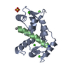


 PDBj
PDBj


 Assembly
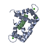


 Mass: 40.078 Da / Num. of mol.: 4 / Source method: obtained synthetically / Formula: Ca
 Mass: 18.015 Da / Num. of mol.: 122 / Source method: isolated from a natural source / Formula: H2O
 Sample preparation
 / Beamline: 21-ID-G / Wavelength: 0.97857 Å
 Processing