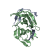
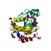
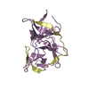
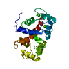
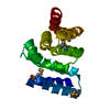
[English] 日本語
Yorodumi
- PDB-5jqa: CaM:RM20 complex -

+
Open data


ID or keywords:

Loading...

-
Basic information

Entry
Database: PDB / ID: 5jqa
TitleCaM:RM20 complex
Components
  • Calmodulin
  • Myosin light chain kinase, smooth muscle
KeywordsCALCIUM BINDING PROTEIN/PROTEIN BINDING / calmodulin / calcium signal transduction / protein kinase / myosin light chain kinase / CALCIUM BINDING PROTEIN-PROTEIN BINDING complex
Function / homology
Function and homology information


aorta smooth muscle tissue morphogenesis / tonic smooth muscle contraction / myosin-light-chain kinase / myosin light chain kinase activity / cellular hypotonic response / : / bleb assembly / establishment of protein localization to mitochondrial membrane / type 3 metabotropic glutamate receptor binding / positive regulation of calcium ion transport ...aorta smooth muscle tissue morphogenesis / tonic smooth muscle contraction / myosin-light-chain kinase / myosin light chain kinase activity / cellular hypotonic response / : / bleb assembly / establishment of protein localization to mitochondrial membrane / type 3 metabotropic glutamate receptor binding / positive regulation of calcium ion transport / CaM pathway / Cam-PDE 1 activation / Sodium/Calcium exchangers / regulation of synaptic vesicle endocytosis / Calmodulin induced events / Reduction of cytosolic Ca++ levels / CREB1 phosphorylation through the activation of CaMKII/CaMKK/CaMKIV cascasde / Activation of Ca-permeable Kainate Receptor / Loss of phosphorylation of MECP2 at T308 / CREB1 phosphorylation through the activation of Adenylate Cyclase / PKA activation / negative regulation of high voltage-gated calcium channel activity / CaMK IV-mediated phosphorylation of CREB / positive regulation of cyclic-nucleotide phosphodiesterase activity / Glycogen breakdown (glycogenolysis) / organelle localization by membrane tethering / negative regulation of calcium ion export across plasma membrane / regulation of synaptic vesicle exocytosis / CLEC7A (Dectin-1) induces NFAT activation / regulation of cardiac muscle cell action potential / autophagosome membrane docking / mitochondrion-endoplasmic reticulum membrane tethering / Activation of RAC1 downstream of NMDARs / positive regulation of wound healing / response to corticosterone / nitric-oxide synthase binding / positive regulation of ryanodine-sensitive calcium-release channel activity / regulation of cell communication by electrical coupling involved in cardiac conduction / Negative regulation of NMDA receptor-mediated neuronal transmission / negative regulation of peptidyl-threonine phosphorylation / Synthesis of IP3 and IP4 in the cytosol / Unblocking of NMDA receptors, glutamate binding and activation / Phase 0 - rapid depolarisation / protein phosphatase activator activity / RHO GTPases activate PAKs / cleavage furrow / positive regulation of phosphoprotein phosphatase activity / Ion transport by P-type ATPases / Long-term potentiation / Uptake and function of anthrax toxins / adenylate cyclase binding / Regulation of MECP2 expression and activity / Calcineurin activates NFAT / catalytic complex / DARPP-32 events / detection of calcium ion / smooth muscle contraction / regulation of cardiac muscle contraction / negative regulation of ryanodine-sensitive calcium-release channel activity / Smooth Muscle Contraction / calcium channel inhibitor activity / RHO GTPases activate IQGAPs / cellular response to interferon-beta / positive regulation of DNA binding / regulation of cardiac muscle contraction by regulation of the release of sequestered calcium ion / Protein methylation / phosphatidylinositol 3-kinase binding / eNOS activation / Activation of AMPK downstream of NMDARs / regulation of release of sequestered calcium ion into cytosol by sarcoplasmic reticulum / enzyme regulator activity / stress fiber / Tetrahydrobiopterin (BH4) synthesis, recycling, salvage and regulation / positive regulation of protein dephosphorylation / regulation of calcium-mediated signaling / Ion homeostasis / titin binding / regulation of ryanodine-sensitive calcium-release channel activity / voltage-gated potassium channel complex / positive regulation of protein autophosphorylation / sperm midpiece / calcium channel complex / response to amphetamine / activation of adenylate cyclase activity / substantia nigra development / adenylate cyclase activator activity / Ras activation upon Ca2+ influx through NMDA receptor / nitric-oxide synthase regulator activity / regulation of heart rate / sarcomere / protein serine/threonine kinase activator activity / FCERI mediated Ca+2 mobilization / FCGR3A-mediated IL10 synthesis / VEGFR2 mediated vascular permeability / positive regulation of peptidyl-threonine phosphorylation / regulation of cytokinesis / Antigen activates B Cell Receptor (BCR) leading to generation of second messengers / VEGFR2 mediated cell proliferation / positive regulation of nitric-oxide synthase activity / Translocation of SLC2A4 (GLUT4) to the plasma membrane
Similarity search - Function
Myosin Light Chain Kinase 1, Kinase domain / Unstructured linker between I-set domains 2 and 3 on MYLCK / Immunoglobulin I-set / Immunoglobulin I-set domain / Fibronectin type III domain / Fibronectin type 3 domain / EF-hand domain pair / Immunoglobulin subtype 2 / Immunoglobulin C-2 Type / Fibronectin type-III domain profile. ...Myosin Light Chain Kinase 1, Kinase domain / Unstructured linker between I-set domains 2 and 3 on MYLCK / Immunoglobulin I-set / Immunoglobulin I-set domain / Fibronectin type III domain / Fibronectin type 3 domain / EF-hand domain pair / Immunoglobulin subtype 2 / Immunoglobulin C-2 Type / Fibronectin type-III domain profile. / Fibronectin type III / Fibronectin type III superfamily / EF-hand, calcium binding motif / EF-Hand 1, calcium-binding site / EF-hand calcium-binding domain. / EF-hand calcium-binding domain profile. / EF-hand domain / EF-hand domain pair / Immunoglobulin subtype / Immunoglobulin / Ig-like domain profile. / Immunoglobulin-like domain / Serine/threonine-protein kinase, active site / Serine/Threonine protein kinases active-site signature. / Immunoglobulin-like domain superfamily / Protein kinase domain / Serine/Threonine protein kinases, catalytic domain / Protein kinase, ATP binding site / Protein kinases ATP-binding region signature. / Immunoglobulin-like fold / Protein kinase domain profile. / Protein kinase domain / Protein kinase-like domain superfamily
Similarity search - Domain/homology
Calmodulin-1 / Calmodulin-3 / Myosin light chain kinase, smooth muscle
Similarity search - Component
Biological speciesHomo sapiens (human)
MethodX-RAY DIFFRACTION / SYNCHROTRON / MOLECULAR REPLACEMENT / Resolution: 1.8 Å
AuthorsTokars, V.L. / Minasov, G. / Anderson, W.F. / Watterson, D.M.
CitationJournal: To be Published
Title: CaM:RM20 complex
Authors: Tokars, V.L. / Minasov, G. / Anderson, W.F. / Watterson, D.M.
History
DepositionMay 4, 2016Deposition site: RCSB / Processing site: RCSB
Revision 1.0Oct 11, 2017Provider: repository / Type: Initial release
Revision 1.1Sep 27, 2023Group: Data collection / Database references ...Data collection / Database references / Derived calculations / Refinement description
Category: chem_comp_atom / chem_comp_bond ...chem_comp_atom / chem_comp_bond / database_2 / pdbx_initial_refinement_model / struct_conn / struct_conn_type
Item: _database_2.pdbx_DOI / _database_2.pdbx_database_accession ..._database_2.pdbx_DOI / _database_2.pdbx_database_accession / _struct_conn.conn_type_id / _struct_conn.id / _struct_conn.pdbx_dist_value / _struct_conn.pdbx_leaving_atom_flag / _struct_conn.ptnr1_auth_asym_id / _struct_conn.ptnr1_auth_comp_id / _struct_conn.ptnr1_auth_seq_id / _struct_conn.ptnr1_label_asym_id / _struct_conn.ptnr1_label_atom_id / _struct_conn.ptnr1_label_comp_id / _struct_conn.ptnr1_label_seq_id / _struct_conn.ptnr2_auth_asym_id / _struct_conn.ptnr2_auth_comp_id / _struct_conn.ptnr2_auth_seq_id / _struct_conn.ptnr2_label_asym_id / _struct_conn.ptnr2_label_atom_id / _struct_conn.ptnr2_label_comp_id / _struct_conn.ptnr2_label_seq_id / _struct_conn_type.id

-
Structure visualization

Structure viewerMolecule:
MolmilJmol/JSmol

Downloads & links

-
Assembly

Deposited unit
A: Calmodulin
B: Myosin light chain kinase, smooth muscle
hetero molecules


Theoretical massNumber of molelcules
Total (without water)19,3336
Polymers19,1722
Non-polymers1604
Water2,792155
1


  • Idetical with deposited unit
  • defined by author&software
TypeNameSymmetry operationNumber
identity operation1_555x,y,z1
Buried area3630 Å2
ΔGint-73 kcal/mol
Surface area8770 Å2
MethodPISA
Unit cell
Length a, b, c (Å)29.016, 57.048, 45.068
Angle α, β, γ (deg.)90.00, 98.17, 90.00
Int Tables number4
Space group name H-MP1211

-
Components

#1: Protein Calmodulin / CaM


Mass: 16852.545 Da / Num. of mol.: 1
Source method: isolated from a genetically manipulated source
Source: (gene. exp.) Homo sapiens (human)
Gene: CALM1, CALM, CAM, CAM1, CALM2, CAM2, CAMB, CALM3, CALML2, CAM3, CAMC, CAMIII
Production host: Escherichia coli (E. coli) / References: UniProt: P62158, UniProt: P0DP23*PLUS
#2: Protein/peptide Myosin light chain kinase, smooth muscle / smMLCK / Kinase-related protein / KRP / Telokin


Mass: 2319.760 Da / Num. of mol.: 1 / Fragment: UNP residues 1691-1710 / Source method: obtained synthetically / Source: (synth.) Homo sapiens (human) / References: UniProt: Q15746
#3: Chemical
ChemComp-CA / CALCIUM ION


Mass: 40.078 Da / Num. of mol.: 4 / Source method: obtained synthetically / Formula: Ca
#4: Water ChemComp-HOH / water


Mass: 18.015 Da / Num. of mol.: 155 / Source method: isolated from a natural source / Formula: H2O

-
Experimental details

-
Experiment

ExperimentMethod: X-RAY DIFFRACTION / Number of used crystals: 1

-
Sample preparation

CrystalDensity Matthews: 1.93 Å3/Da / Density % sol: 36.13 %
Crystal growTemperature: 295 K / Method: vapor diffusion, sitting drop / Details: 0.1 M sodium acetate, pH 4.6, 25% w/v PEG4000

-
Data collection

DiffractionMean temperature: 100 K
Diffraction sourceSource: SYNCHROTRON / Site: APS / Beamline: 21-ID-F / Wavelength: 0.97872 Å
DetectorType: MARMOSAIC 225 mm CCD / Detector: CCD / Date: Nov 24, 2014
RadiationMonochromator: diamond(111) / Protocol: SINGLE WAVELENGTH / Monochromatic (M) / Laue (L): M / Scattering type: x-ray
Radiation wavelengthWavelength: 0.97872 Å / Relative weight: 1
ReflectionResolution: 1.72→50 Å / Num. obs: 15660 / % possible obs: 95.5 % / Redundancy: 96.6 % / Net I/σ(I): 14.1
Reflection shellHighest resolution: 1.72 Å

-
Processing

Software
NameVersionClassification
REFMAC5.8.0049refinement
HKL-2000data reduction
HKL-2000data scaling
PHASERphasing
RefinementMethod to determine structure: MOLECULAR REPLACEMENT
Starting model: PDB entry 2O5G
Resolution: 1.8→28.72 Å / Cor.coef. Fo:Fc: 0.968 / Cor.coef. Fo:Fc free: 0.952 / SU B: 5.796 / SU ML: 0.092 / Cross valid method: THROUGHOUT / ESU R: 0.142 / ESU R Free: 0.123 / Details: HYDROGENS HAVE BEEN ADDED IN THE RIDING POSITIONS
RfactorNum. reflection% reflectionSelection details
Rfree0.18845 658 4.9 %RANDOM
Rwork0.1534 ---
obs0.15515 12699 98.45 %-
Solvent computationIon probe radii: 0.8 Å / Shrinkage radii: 0.8 Å / VDW probe radii: 1.2 Å
Displacement parametersBiso mean: 25.522 Å2
Baniso -1Baniso -2Baniso -3
1-0.26 Å20 Å20.18 Å2
2---0.24 Å20 Å2
3----0.06 Å2
Refinement stepCycle: LAST / Resolution: 1.8→28.72 Å
ProteinNucleic acidLigandSolventTotal
Num. atoms1323 0 4 155 1482
Refine LS restraints
Refine-IDTypeDev idealDev ideal targetNumber
X-RAY DIFFRACTIONr_bond_refined_d0.010.0191464
X-RAY DIFFRACTIONr_bond_other_d0.0010.021380
X-RAY DIFFRACTIONr_angle_refined_deg1.3571.9611971
X-RAY DIFFRACTIONr_angle_other_deg0.81933195
X-RAY DIFFRACTIONr_dihedral_angle_1_deg3.1615187
X-RAY DIFFRACTIONr_dihedral_angle_2_deg27.06525.43281
X-RAY DIFFRACTIONr_dihedral_angle_3_deg10.64215293
X-RAY DIFFRACTIONr_dihedral_angle_4_deg15.7051512
X-RAY DIFFRACTIONr_chiral_restr0.0950.2209
X-RAY DIFFRACTIONr_gen_planes_refined0.0220.021725
X-RAY DIFFRACTIONr_gen_planes_other0.0180.02327
X-RAY DIFFRACTIONr_nbd_refined
X-RAY DIFFRACTIONr_nbd_other
X-RAY DIFFRACTIONr_nbtor_refined
X-RAY DIFFRACTIONr_nbtor_other
X-RAY DIFFRACTIONr_xyhbond_nbd_refined
X-RAY DIFFRACTIONr_xyhbond_nbd_other
X-RAY DIFFRACTIONr_metal_ion_refined
X-RAY DIFFRACTIONr_metal_ion_other
X-RAY DIFFRACTIONr_symmetry_vdw_refined
X-RAY DIFFRACTIONr_symmetry_vdw_other
X-RAY DIFFRACTIONr_symmetry_hbond_refined
X-RAY DIFFRACTIONr_symmetry_hbond_other
X-RAY DIFFRACTIONr_symmetry_metal_ion_refined
X-RAY DIFFRACTIONr_symmetry_metal_ion_other
X-RAY DIFFRACTIONr_mcbond_it2.0871.866725
X-RAY DIFFRACTIONr_mcbond_other2.0751.857723
X-RAY DIFFRACTIONr_mcangle_it3.2572.758919
X-RAY DIFFRACTIONr_mcangle_other3.2572.763920
X-RAY DIFFRACTIONr_scbond_it2.7422.256739
X-RAY DIFFRACTIONr_scbond_other2.742.259740
X-RAY DIFFRACTIONr_scangle_it
X-RAY DIFFRACTIONr_scangle_other4.4813.2321053
X-RAY DIFFRACTIONr_long_range_B_refined7.06615.9471972
X-RAY DIFFRACTIONr_long_range_B_other7.00415.4781907
X-RAY DIFFRACTIONr_rigid_bond_restr
X-RAY DIFFRACTIONr_sphericity_free
X-RAY DIFFRACTIONr_sphericity_bonded
LS refinement shellResolution: 1.8→1.847 Å / Total num. of bins used: 20
RfactorNum. reflection% reflection
Rfree0.256 52 -
Rwork0.219 910 -
obs--97.07 %
Refinement TLS params.

Method: refined / Refine-ID: X-RAY DIFFRACTION

IDL112)L122)L132)L222)L232)L332)S11 (Å °)S12 (Å °)S13 (Å °)S21 (Å °)S22 (Å °)S23 (Å °)S31 (Å °)S32 (Å °)S33 (Å °)T112)T122)T132)T222)T232)T332)Origin x (Å)Origin y (Å)Origin z (Å)
10.5434-0.33930.76990.4639-0.40131.1206-0.0695-0.0729-0.01080.05070.06990.0122-0.0978-0.0919-0.00030.01640.0255-0.00420.05190.00440.0371-6.92661.2518-7.7556
20.90140.0534-0.070.5276-0.12960.1594-0.0088-0.03680.0725-0.0134-0.0003-0.00780.0346-0.01340.00910.0167-0.0057-0.01260.0459-0.00340.0337-1.3731-17.5265-14.1849
32.8862-1.8709-1.60741.46570.68551.401-0.0578-0.01370.0596-0.00040.054-0.06990.0872-0.06580.00380.0113-0.0048-0.00910.0573-0.00780.0402-3.2795-7.374-7.1853
Refinement TLS group
IDRefine-IDRefine TLS-IDAuth asym-IDAuth seq-ID
1X-RAY DIFFRACTION1A2 - 77
2X-RAY DIFFRACTION1A401 - 402
3X-RAY DIFFRACTION2A78 - 148
4X-RAY DIFFRACTION2A403 - 404
5X-RAY DIFFRACTION3B1 - 21

+
About Yorodumi

-
News

-
Feb 9, 2022. New format data for meta-information of EMDB entries

New format data for meta-information of EMDB entries

  • Version 3 of the EMDB header file is now the official format.
  • The previous official version 1.9 will be removed from the archive.

Related info.:EMDB header

External links:wwPDB to switch to version 3 of the EMDB data model

-
Aug 12, 2020. Covid-19 info

Covid-19 info

URL: https://pdbjlvh1.pdbj.org/emnavi/covid19.php

New page: Covid-19 featured information page in EM Navigator.

Related info.:Covid-19 info / Mar 5, 2020. Novel coronavirus structure data

+
Mar 5, 2020. Novel coronavirus structure data

Novel coronavirus structure data

Related info.:Yorodumi Speices / Aug 12, 2020. Covid-19 info

External links:COVID-19 featured content - PDBj / Molecule of the Month (242):Coronavirus Proteases

+
Jan 31, 2019. EMDB accession codes are about to change! (news from PDBe EMDB page)

EMDB accession codes are about to change! (news from PDBe EMDB page)

  • The allocation of 4 digits for EMDB accession codes will soon come to an end. Whilst these codes will remain in use, new EMDB accession codes will include an additional digit and will expand incrementally as the available range of codes is exhausted. The current 4-digit format prefixed with “EMD-” (i.e. EMD-XXXX) will advance to a 5-digit format (i.e. EMD-XXXXX), and so on. It is currently estimated that the 4-digit codes will be depleted around Spring 2019, at which point the 5-digit format will come into force.
  • The EM Navigator/Yorodumi systems omit the EMD- prefix.

Related info.:Q: What is EMD? / ID/Accession-code notation in Yorodumi/EM Navigator

External links:EMDB Accession Codes are Changing Soon! / Contact to PDBj

+
Jul 12, 2017. Major update of PDB

Major update of PDB

  • wwPDB released updated PDB data conforming to the new PDBx/mmCIF dictionary.
  • This is a major update changing the version number from 4 to 5, and with Remediation, in which all the entries are updated.
  • In this update, many items about electron microscopy experimental information are reorganized (e.g. em_software).
  • Now, EM Navigator and Yorodumi are based on the updated data.

External links:wwPDB Remediation / Enriched Model Files Conforming to OneDep Data Standards Now Available in the PDB FTP Archive

-
Yorodumi

Thousand views of thousand structures

  • Yorodumi is a browser for structure data from EMDB, PDB, SASBDB, etc.
  • This page is also the successor to EM Navigator detail page, and also detail information page/front-end page for Omokage search.
  • The word "yorodu" (or yorozu) is an old Japanese word meaning "ten thousand". "mi" (miru) is to see.

Related info.:EMDB / PDB / SASBDB / Comparison of 3 databanks / Yorodumi Search / Aug 31, 2016. New EM Navigator & Yorodumi / Yorodumi Papers / Jmol/JSmol / Function and homology information / Changes in new EM Navigator and Yorodumi

Read more