[English] 日本語
Yorodumi
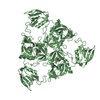
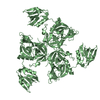
- PDB-5jd8: Crystal structure of the serine endoprotease from Yersinia pestis -

+
Open data


ID or keywords:

Loading...

-
Basic information

Entry
Database: PDB / ID: 5jd8
TitleCrystal structure of the serine endoprotease from Yersinia pestis
ComponentsPeriplasmic serine peptidase DegS
KeywordsHYDROLASE / DegS / Structural Genomics / Center for Structural Genomics of Infectious Diseases / CSGID
Function / homology
Function and homology information


Hydrolases; Acting on peptide bonds (peptidases); Serine endopeptidases / serine-type endopeptidase activity
Similarity search - Function
Peptidase S1C, DegS / PDZ domain / Peptidase S1C / Trypsin-like peptidase domain / Domain present in PSD-95, Dlg, and ZO-1/2. / PDZ domain / PDZ superfamily / Trypsin-like serine proteases / Thrombin, subunit H / Peptidase S1, PA clan, chymotrypsin-like fold ...Peptidase S1C, DegS / PDZ domain / Peptidase S1C / Trypsin-like peptidase domain / Domain present in PSD-95, Dlg, and ZO-1/2. / PDZ domain / PDZ superfamily / Trypsin-like serine proteases / Thrombin, subunit H / Peptidase S1, PA clan, chymotrypsin-like fold / Peptidase S1, PA clan / Beta Barrel / Mainly Beta
Similarity search - Domain/homology
DI(HYDROXYETHYL)ETHER / : / Protease
Similarity search - Component
Biological speciesYersinia pestis CO92 (bacteria)
MethodX-RAY DIFFRACTION / SYNCHROTRON / MOLECULAR REPLACEMENT / Resolution: 1.85 Å
AuthorsFilippova, E.V. / Wawrzsak, Z. / Sandoval, J. / Skarina, T. / Grimshaw, S. / Savchenko, A. / Anderson, W.F. / Center for Structural Genomics of Infectious Diseases (CSGID)
CitationJournal: To Be Published
Title: Crystal structure of the serine endoprotease from Yersinia pestis
Authors: Filippova, E.V. / Wawrzsak, Z. / Sandoval, J. / Skarina, T. / Grimshaw, S. / Savchenko, A. / Anderson, W.F. / Center for Structural Genomics of Infectious Diseases (CSGID)
History
DepositionApr 15, 2016Deposition site: RCSB / Processing site: RCSB
Revision 1.0May 4, 2016Provider: repository / Type: Initial release
Revision 1.1Nov 22, 2017Group: Derived calculations / Refinement description / Category: pdbx_struct_oper_list / software / Item: _pdbx_struct_oper_list.symmetry_operation
Revision 1.2Sep 27, 2023Group: Data collection / Database references / Refinement description
Category: chem_comp_atom / chem_comp_bond ...chem_comp_atom / chem_comp_bond / database_2 / pdbx_initial_refinement_model
Item: _database_2.pdbx_DOI / _database_2.pdbx_database_accession

-
Structure visualization

Structure viewerMolecule:
MolmilJmol/JSmol

Downloads & links

-
Assembly

Deposited unit
A: Periplasmic serine peptidase DegS
hetero molecules


Theoretical massNumber of molelcules
Total (without water)35,7784
Polymers35,3551
Non-polymers4243
Water2,774154
1
A: Periplasmic serine peptidase DegS
hetero molecules

A: Periplasmic serine peptidase DegS
hetero molecules

A: Periplasmic serine peptidase DegS
hetero molecules


Theoretical massNumber of molelcules
Total (without water)107,33512
Polymers106,0643
Non-polymers1,2719
Water543
TypeNameSymmetry operationNumber
identity operation1_555x,y,z1
crystal symmetry operation2_665-y+1,x-y+1,z1
crystal symmetry operation3_565-x+y,-x+1,z1
Buried area8430 Å2
ΔGint-77 kcal/mol
Surface area37040 Å2
MethodPISA
Unit cell
Length a, b, c (Å)104.418, 104.418, 76.015
Angle α, β, γ (deg.)90.00, 90.00, 120.00
Int Tables number146
Space group name H-MH3
Components on special symmetry positions
IDModelComponents
11A-403-

SO4

21A-553-

HOH

-
Components

#1: Protein Periplasmic serine peptidase DegS


Mass: 35354.785 Da / Num. of mol.: 1 / Fragment: UNP residues 29-362
Source method: isolated from a genetically manipulated source
Source: (gene. exp.) Yersinia pestis CO92 (bacteria) / Gene: degS, AK38_3169 / Plasmid: pMCSG53 / Production host: Escherichia coli (E. coli) / Strain (production host): BL21(DE3)-Magic / References: UniProt: A0A0B6NK33, UniProt: A0A5P8YL96*PLUS
#2: Chemical ChemComp-CXS / 3-CYCLOHEXYL-1-PROPYLSULFONIC ACID / CAPS (buffer)


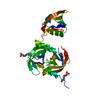
Mass: 221.317 Da / Num. of mol.: 1 / Source method: obtained synthetically / Formula: C9H19NO3S / Comment: pH buffer*YM
#3: Chemical ChemComp-PEG / DI(HYDROXYETHYL)ETHER / Diethylene glycol


Mass: 106.120 Da / Num. of mol.: 1 / Source method: obtained synthetically / Formula: C4H10O3
#4: Chemical ChemComp-SO4 / SULFATE ION / Sulfate


Mass: 96.063 Da / Num. of mol.: 1 / Source method: obtained synthetically / Formula: SO4
#5: Water ChemComp-HOH / water / Water


Mass: 18.015 Da / Num. of mol.: 154 / Source method: isolated from a natural source / Formula: H2O

-
Experimental details

-
Experiment

ExperimentMethod: X-RAY DIFFRACTION / Number of used crystals: 1

-
Sample preparation

CrystalDensity Matthews: 2.26 Å3/Da / Density % sol: 45.47 %
Crystal growTemperature: 293 K / Method: vapor diffusion, hanging drop / pH: 5.6
Details: 20% PEG 4000, 10% Isopropane, 50 mM CAPSO buffer pH 11, 0.1 M Sodium citrate pH 5.6, 5% P200 paratone-N oil

-
Data collection

DiffractionMean temperature: 100 K
Diffraction sourceSource: SYNCHROTRON / Site: APS / Beamline: 21-ID-F / Wavelength: 0.97872 Å
DetectorType: MARMOSAIC 225 mm CCD / Detector: CCD / Date: Feb 3, 2016 / Details: beryllium lenses
RadiationMonochromator: C(111) / Protocol: SINGLE WAVELENGTH / Monochromatic (M) / Laue (L): M / Scattering type: x-ray
Radiation wavelengthWavelength: 0.97872 Å / Relative weight: 1
ReflectionResolution: 1.85→30 Å / Num. obs: 26479 / % possible obs: 100 % / Redundancy: 7.1 % / Rmerge(I) obs: 0.1 / Net I/σ(I): 15.8
Reflection shellResolution: 1.85→1.88 Å / Redundancy: 6.9 % / Rmerge(I) obs: 0.65 / Mean I/σ(I) obs: 3.3 / % possible all: 100

-
Processing

Software
NameVersionClassification
REFMAC5.8.0135refinement
HKL-2000data scaling
PHENIXphasing
Cootmodel building
RefinementMethod to determine structure: MOLECULAR REPLACEMENT
Starting model: 4RQY
Resolution: 1.85→30 Å / Cor.coef. Fo:Fc: 0.966 / Cor.coef. Fo:Fc free: 0.946 / SU B: 4.311 / SU ML: 0.064 / Cross valid method: THROUGHOUT / ESU R: 0.103 / ESU R Free: 0.102 / Stereochemistry target values: MAXIMUM LIKELIHOOD / Details: HYDROGENS HAVE BEEN ADDED IN THE RIDING POSITIONS
RfactorNum. reflection% reflectionSelection details
Rfree0.18269 1292 4.9 %RANDOM
Rwork0.14831 ---
obs0.14999 25187 99.91 %-
Solvent computationIon probe radii: 0.8 Å / Shrinkage radii: 0.8 Å / VDW probe radii: 1.2 Å / Solvent model: MASK
Displacement parametersBiso mean: 46.116 Å2
Baniso -1Baniso -2Baniso -3
1--0.38 Å2-0.19 Å20 Å2
2---0.38 Å20 Å2
3---1.24 Å2
Refinement stepCycle: LAST / Resolution: 1.85→30 Å
ProteinNucleic acidLigandSolventTotal
Num. atoms2017 0 26 154 2197
Refine LS restraints
Refine-IDTypeDev idealDev ideal targetNumber
X-RAY DIFFRACTIONr_bond_refined_d0.0210.0192081
X-RAY DIFFRACTIONr_bond_other_d0.0020.022115
X-RAY DIFFRACTIONr_angle_refined_deg2.0031.9852828
X-RAY DIFFRACTIONr_angle_other_deg1.04834843
X-RAY DIFFRACTIONr_dihedral_angle_1_deg7.2365271
X-RAY DIFFRACTIONr_dihedral_angle_2_deg37.47525.37580
X-RAY DIFFRACTIONr_dihedral_angle_3_deg15.73815354
X-RAY DIFFRACTIONr_dihedral_angle_4_deg14.8461513
X-RAY DIFFRACTIONr_chiral_restr0.1280.2355
X-RAY DIFFRACTIONr_gen_planes_refined0.010.0212347
X-RAY DIFFRACTIONr_gen_planes_other0.0020.02424
X-RAY DIFFRACTIONr_nbd_refined
X-RAY DIFFRACTIONr_nbd_other
X-RAY DIFFRACTIONr_nbtor_refined
X-RAY DIFFRACTIONr_nbtor_other
X-RAY DIFFRACTIONr_xyhbond_nbd_refined
X-RAY DIFFRACTIONr_xyhbond_nbd_other
X-RAY DIFFRACTIONr_metal_ion_refined
X-RAY DIFFRACTIONr_metal_ion_other
X-RAY DIFFRACTIONr_symmetry_vdw_refined
X-RAY DIFFRACTIONr_symmetry_vdw_other
X-RAY DIFFRACTIONr_symmetry_hbond_refined
X-RAY DIFFRACTIONr_symmetry_hbond_other
X-RAY DIFFRACTIONr_symmetry_metal_ion_refined
X-RAY DIFFRACTIONr_symmetry_metal_ion_other
X-RAY DIFFRACTIONr_mcbond_it2.3333.2341093
X-RAY DIFFRACTIONr_mcbond_other2.3313.2331092
X-RAY DIFFRACTIONr_mcangle_it3.8094.8151361
X-RAY DIFFRACTIONr_mcangle_other3.8084.8171362
X-RAY DIFFRACTIONr_scbond_it2.8893.643988
X-RAY DIFFRACTIONr_scbond_other2.8813.627984
X-RAY DIFFRACTIONr_scangle_it
X-RAY DIFFRACTIONr_scangle_other4.6135.2931462
X-RAY DIFFRACTIONr_long_range_B_refined7.96825.6242188
X-RAY DIFFRACTIONr_long_range_B_other7.96625.6312189
X-RAY DIFFRACTIONr_rigid_bond_restr
X-RAY DIFFRACTIONr_sphericity_free
X-RAY DIFFRACTIONr_sphericity_bonded
LS refinement shellResolution: 1.848→1.896 Å / Total num. of bins used: 20
RfactorNum. reflection% reflection
Rfree0.2 91 -
Rwork0.201 1871 -
obs--99.09 %
Refinement TLS params.

Method: refined / Refine-ID: X-RAY DIFFRACTION

IDL112)L122)L132)L222)L232)L332)S11 (Å °)S12 (Å °)S13 (Å °)S21 (Å °)S22 (Å °)S23 (Å °)S31 (Å °)S32 (Å °)S33 (Å °)T112)T122)T132)T222)T232)T332)Origin x (Å)Origin y (Å)Origin z (Å)
10.6618-0.9985-0.42221.65350.56670.62750.02690.01320.0847-0.0401-0.0615-0.22940.0304-0.00330.03460.04490.01770.03520.05820.01170.129520.700747.065650.4939
20.9328-0.3613-0.08210.39450.03660.08660.01290.0397-0.1135-0.0398-0.019-0.0138-0.0046-0.00390.00610.06950.00860.01090.05810.00660.09017.210849.353553.9369
31.71290.6593-1.14710.73040.17061.5592-0.2290.1031-0.29190.0298-0.0235-0.07310.3011-0.17170.25260.1153-0.00680.07280.0315-0.0080.20810.953229.217249.0316
41.8574-1.07562.36756.53031.62164.65320.2295-0.0797-0.3372-0.2508-0.12170.62360.35-0.3537-0.10780.1449-0.16960.05250.2412-0.14440.1847-3.274315.748739.0372
510.2623-5.71640.22315.61931.6171.35820.0425-0.2805-0.11010.1902-0.12190.05460.27-0.30070.07940.1491-0.12940.08630.1241-0.07140.07592.278521.22447.5389
612.27440.10955.24830.29730.0630.0736-0.0238-0.34470.1557-0.114-0.69020.0572-0.11960.04040.1592-0.20960.00990.3536-0.13330.2497-6.77138.28442.0678
74.47553.12640.37146.89921.06350.34420.08620.7828-0.39540.47810.0299-0.04220.17370.1044-0.11610.18690.0306-0.00060.2864-0.18810.16148.261916.176642.7082
Refinement TLS group
IDRefine-IDRefine TLS-IDAuth asym-IDAuth seq-ID
1X-RAY DIFFRACTION1A43 - 134
2X-RAY DIFFRACTION2A135 - 234
3X-RAY DIFFRACTION3A235 - 263
4X-RAY DIFFRACTION4A280 - 311
5X-RAY DIFFRACTION5A312 - 329
6X-RAY DIFFRACTION6A330 - 343
7X-RAY DIFFRACTION7A344 - 351

+
About Yorodumi

-
News

-
Feb 9, 2022. New format data for meta-information of EMDB entries

New format data for meta-information of EMDB entries

  • Version 3 of the EMDB header file is now the official format.
  • The previous official version 1.9 will be removed from the archive.

Related info.:EMDB header

External links:wwPDB to switch to version 3 of the EMDB data model

-
Aug 12, 2020. Covid-19 info

Covid-19 info

URL: https://pdbj.org/emnavi/covid19.php

New page: Covid-19 featured information page in EM Navigator.

Related info.:Covid-19 info / Mar 5, 2020. Novel coronavirus structure data

+
Mar 5, 2020. Novel coronavirus structure data

Novel coronavirus structure data

Related info.:Yorodumi Speices / Aug 12, 2020. Covid-19 info

External links:COVID-19 featured content - PDBj / Molecule of the Month (242):Coronavirus Proteases

+
Jan 31, 2019. EMDB accession codes are about to change! (news from PDBe EMDB page)

EMDB accession codes are about to change! (news from PDBe EMDB page)

  • The allocation of 4 digits for EMDB accession codes will soon come to an end. Whilst these codes will remain in use, new EMDB accession codes will include an additional digit and will expand incrementally as the available range of codes is exhausted. The current 4-digit format prefixed with “EMD-” (i.e. EMD-XXXX) will advance to a 5-digit format (i.e. EMD-XXXXX), and so on. It is currently estimated that the 4-digit codes will be depleted around Spring 2019, at which point the 5-digit format will come into force.
  • The EM Navigator/Yorodumi systems omit the EMD- prefix.

Related info.:Q: What is EMD? / ID/Accession-code notation in Yorodumi/EM Navigator

External links:EMDB Accession Codes are Changing Soon! / Contact to PDBj

+
Jul 12, 2017. Major update of PDB

Major update of PDB

  • wwPDB released updated PDB data conforming to the new PDBx/mmCIF dictionary.
  • This is a major update changing the version number from 4 to 5, and with Remediation, in which all the entries are updated.
  • In this update, many items about electron microscopy experimental information are reorganized (e.g. em_software).
  • Now, EM Navigator and Yorodumi are based on the updated data.

External links:wwPDB Remediation / Enriched Model Files Conforming to OneDep Data Standards Now Available in the PDB FTP Archive

-
Yorodumi

Thousand views of thousand structures

  • Yorodumi is a browser for structure data from EMDB, PDB, SASBDB, etc.
  • This page is also the successor to EM Navigator detail page, and also detail information page/front-end page for Omokage search.
  • The word "yorodu" (or yorozu) is an old Japanese word meaning "ten thousand". "mi" (miru) is to see.

Related info.:EMDB / PDB / SASBDB / Comparison of 3 databanks / Yorodumi Search / Aug 31, 2016. New EM Navigator & Yorodumi / Yorodumi Papers / Jmol/JSmol / Function and homology information / Changes in new EM Navigator and Yorodumi

Read more