 Movie
Movie Controller


+ Open data

- Basic information
| Entry | Database: PDB / ID: 5j84 | |||||||||
|---|---|---|---|---|---|---|---|---|---|---|
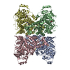
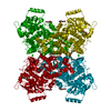
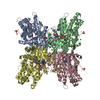
| Title | Crystal structure of L-arabinonate dehydratase in holo-form | |||||||||
 Components Components | Dihydroxy-acid dehydratase | |||||||||
 Keywords Keywords | LYASE / L-arabinonate dehydratase / L-arabonate dehydratase / pentonate dehydratase / 2Fe2S cluster | |||||||||
| Function / homology |  Function and homology information Function and homology informationL-arabinonate dehydratase / D-fuconate dehydratase / D-fuconate dehydratase activity / L-arabinonate dehydratase activity / galactonate dehydratase / galactonate dehydratase activity / arabinose catabolic process / 2 iron, 2 sulfur cluster binding / metal ion binding Similarity search - Function | |||||||||
| Biological species |  Rhizobium leguminosarum bv. trifolii (bacteria) Rhizobium leguminosarum bv. trifolii (bacteria) | |||||||||
| Method |  X-RAY DIFFRACTION / SYNCHROTRON / MOLECULAR REPLACEMENT / Resolution: 2.4 Å X-RAY DIFFRACTION / SYNCHROTRON / MOLECULAR REPLACEMENT / Resolution: 2.4 Å | |||||||||
 Authors Authors | Rahman, M.M. / Rouvinen, J. / Hakulinen, N. | |||||||||
| Funding support |  Finland, 2items Finland, 2items
| |||||||||
 Citation Citation | Journal: ACS Chem. Biol. / Year: 2017 Title: The Crystal Structure of a Bacterial l-Arabinonate Dehydratase Contains a [2Fe-2S] Cluster. Authors: Rahman, M.M. / Andberg, M. / Thangaraj, S.K. / Parkkinen, T. / Penttila, M. / Janis, J. / Koivula, A. / Rouvinen, J. / Hakulinen, N. | |||||||||
| History |
|
- Structure visualization
| Structure viewer | Molecule: MolmilJmol/JSmol |
|---|
- Downloads & links
-Download
| PDBx/mmCIF format | 5j84.cif.gz | 884.5 KB | Display | PDBx/mmCIF format |
|---|---|---|---|---|
| PDB format | pdb5j84.ent.gz | 736.8 KB | Display | PDB format |
| PDBx/mmJSON format | 5j84.json.gz | Tree view | PDBx/mmJSON format | |
| Others |  Other downloads Other downloads |
-Validation report
| Summary document | 5j84_validation.pdf.gz | 513.6 KB | Display | wwPDB validaton report |
|---|---|---|---|---|
| Full document | 5j84_full_validation.pdf.gz | 546.6 KB | Display | |
| Data in XML | 5j84_validation.xml.gz | 168 KB | Display | |
| Data in CIF | 5j84_validation.cif.gz | 234.9 KB | Display | |
| Arichive directory | https://data.pdbj.org/pub/pdb/validation_reports/j8/5j84ftp://data.pdbj.org/pub/pdb/validation_reports/j8/5j84 | HTTPS FTP |
-Related structure data
| Related structure data |  5j83C  5j85C 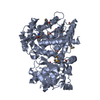 2gp4S S: Starting model for refinement C: citing same article ( |
|---|---|
| Similar structure data |
-Links
 PDBj
PDBj


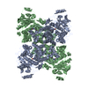
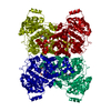
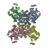
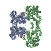
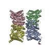
- Assembly
| Deposited unit | 
| ||||||||
|---|---|---|---|---|---|---|---|---|---|
| 1 | 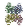
| ||||||||
| 2 | 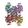
| ||||||||
| Unit cell |
|
-Components
| #1: Protein | Mass: 63858.938 Da / Num. of mol.: 8 Source method: isolated from a genetically manipulated source Details: Gram-negative bacteria Source: (gene. exp.) Rhizobium leguminosarum bv. trifolii (strain WSM2304) (bacteria)Strain: WSM2304 / Gene: Rleg2_2909 / Plasmid: pBAT4 / Production host: #2: Chemical | ChemComp-MG /   Mass: 24.305 Da / Num. of mol.: 8 Mass: 24.305 Da / Num. of mol.: 8Source method: isolated from a genetically manipulated source Formula: Mg / Details: Gram-negative bacteria Source: (gene. exp.) Rhizobium leguminosarum bv. trifolii (bacteria)Gene: 6981653 / Plasmid: pBAT4 / Production host: #3: Chemical | ChemComp-FES /   Mass: 175.820 Da / Num. of mol.: 8 Mass: 175.820 Da / Num. of mol.: 8Source method: isolated from a genetically manipulated source Formula: Fe2S2 / Details: Gram-negative bacteria Source: (gene. exp.) Rhizobium leguminosarum bv. trifolii (bacteria)Gene: 6981653 / Plasmid: pBAT4 / Production host: #4: Water | ChemComp-HOH / |  Mass: 18.015 Da / Num. of mol.: 1771 / Source method: isolated from a natural source / Formula: H2O Mass: 18.015 Da / Num. of mol.: 1771 / Source method: isolated from a natural source / Formula: H2O |
|---|
-Experimental details
-Experiment
| Experiment | Method: X-RAY DIFFRACTION / Number of used crystals: 1 |
|---|
- Sample preparation
| Crystal | Density Matthews: 3.2 Å3/Da / Density % sol: 62 % / Description: 3D-Plate |
|---|---|
| Crystal grow | Temperature: 293 K / Method: vapor diffusion, hanging drop / pH: 7 / Details: 4 M Sodium formate, 0.1 M MES pH 7.0 |
-Data collection
| Diffraction | Mean temperature: 100 K |
|---|---|
| Diffraction source | Source: SYNCHROTRON / Site: ESRF  / Beamline: ID14-2 / Wavelength: 0.97957 Å / Beamline: ID14-2 / Wavelength: 0.97957 Å |
| Detector | Type: ADSC QUANTUM 4 / Detector: CCD / Date: Jun 15, 2012 |
| Radiation | Monochromator: SI(111) / Protocol: SINGLE WAVELENGTH / Monochromatic (M) / Laue (L): M / Scattering type: x-ray |
| Radiation wavelength | Wavelength: 0.97957 Å / Relative weight: 1 |
| Reflection | Resolution: 2.4→40 Å / Num. obs: 246940 / % possible obs: 99.3 % / Redundancy: 3.2 % / Biso Wilson estimate: 43 Å2 / CC1/2: 0.995 / Rsym value: 0.091 / Net I/σ(I): 12.3 |
| Reflection shell | Resolution: 2.4→2.5 Å / Redundancy: 3.1 % / Mean I/σ(I) obs: 2 / % possible all: 99 |
- Processing
| Software |
| |||||||||||||||||||||||||||||||||||||||||||||||||||||||||||||||||||||||||||||||||||||||||||||||||||||||||||||||||||||||||||||||||||||||||||||||||||
|---|---|---|---|---|---|---|---|---|---|---|---|---|---|---|---|---|---|---|---|---|---|---|---|---|---|---|---|---|---|---|---|---|---|---|---|---|---|---|---|---|---|---|---|---|---|---|---|---|---|---|---|---|---|---|---|---|---|---|---|---|---|---|---|---|---|---|---|---|---|---|---|---|---|---|---|---|---|---|---|---|---|---|---|---|---|---|---|---|---|---|---|---|---|---|---|---|---|---|---|---|---|---|---|---|---|---|---|---|---|---|---|---|---|---|---|---|---|---|---|---|---|---|---|---|---|---|---|---|---|---|---|---|---|---|---|---|---|---|---|---|---|---|---|---|---|---|---|---|
| Refinement | Method to determine structure: MOLECULAR REPLACEMENT Starting model: 2GP4 Resolution: 2.4→39.889 Å / Cross valid method: FREE R-VALUE / σ(F): 1.35 / Phase error: 15.35
| |||||||||||||||||||||||||||||||||||||||||||||||||||||||||||||||||||||||||||||||||||||||||||||||||||||||||||||||||||||||||||||||||||||||||||||||||||
| Solvent computation | Shrinkage radii: 0.9 Å / VDW probe radii: 1.11 Å | |||||||||||||||||||||||||||||||||||||||||||||||||||||||||||||||||||||||||||||||||||||||||||||||||||||||||||||||||||||||||||||||||||||||||||||||||||
| Refinement step | Cycle: LAST / Resolution: 2.4→39.889 Å
| |||||||||||||||||||||||||||||||||||||||||||||||||||||||||||||||||||||||||||||||||||||||||||||||||||||||||||||||||||||||||||||||||||||||||||||||||||
| Refine LS restraints |
| |||||||||||||||||||||||||||||||||||||||||||||||||||||||||||||||||||||||||||||||||||||||||||||||||||||||||||||||||||||||||||||||||||||||||||||||||||
| LS refinement shell |
|
