 Movie
Movie Controller


+ Open data

- Basic information
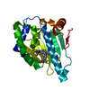
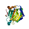
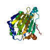
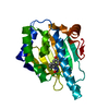
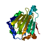
| Entry | Database: PDB / ID: 5iz2 | ||||||
|---|---|---|---|---|---|---|---|
| Title | Crystal structure of the N. clavipes spidroin NTD at pH 6.5 | ||||||
 Components Components |
| ||||||
 Keywords Keywords | STRUCTURAL PROTEIN / spidroin / major ampullate / homodimer | ||||||
| Function / homology | Spidroin, N-terminal domain / Spidroin, N-terminal / Major ampullate spidroin 1, spider silk protein 1, N-term / Spidroin, N-terminal domain superfamily / Enzyme I; Chain A, domain 2 / Orthogonal Bundle / Mainly Alpha / Major ampullate spidroin 1A Function and homology information Function and homology information | ||||||
| Biological species |  Nephila clavipes (spider) Nephila clavipes (spider) | ||||||
| Method |  X-RAY DIFFRACTION / SYNCHROTRON / MOLECULAR REPLACEMENT / Resolution: 2.02 Å X-RAY DIFFRACTION / SYNCHROTRON / MOLECULAR REPLACEMENT / Resolution: 2.02 Å | ||||||
 Authors Authors | Atkison, J.H. / Olsen, S.K. | ||||||
 Citation Citation | Journal: J.Biol.Chem. / Year: 2016 Title: Crystal Structure of the Nephila clavipes Major Ampullate Spidroin 1A N-terminal Domain Reveals Plasticity at the Dimer Interface. Authors: Atkison, J.H. / Parnham, S. / Marcotte, W.R. / Olsen, S.K. | ||||||
| History |
|
- Structure visualization
| Structure viewer | Molecule: MolmilJmol/JSmol |
|---|
- Downloads & links
-Download
| PDBx/mmCIF format | 5iz2.cif.gz | 108.7 KB | Display | PDBx/mmCIF format |
|---|---|---|---|---|
| PDB format | pdb5iz2.ent.gz | 83.5 KB | Display | PDB format |
| PDBx/mmJSON format | 5iz2.json.gz | Tree view | PDBx/mmJSON format | |
| Others |  Other downloads Other downloads |
-Validation report
| Summary document | 5iz2_validation.pdf.gz | 432.6 KB | Display | wwPDB validaton report |
|---|---|---|---|---|
| Full document | 5iz2_full_validation.pdf.gz | 432.7 KB | Display | |
| Data in XML | 5iz2_validation.xml.gz | 11.5 KB | Display | |
| Data in CIF | 5iz2_validation.cif.gz | 15.6 KB | Display | |
| Arichive directory | https://data.pdbj.org/pub/pdb/validation_reports/iz/5iz2ftp://data.pdbj.org/pub/pdb/validation_reports/iz/5iz2 | HTTPS FTP |
-Related structure data
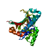
| Related structure data | 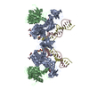 3l2rS S: Starting model for refinement |
|---|---|
| Similar structure data |


-Links
 PDBj
PDBj- Assembly
| Deposited unit | 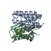
| ||||||||
|---|---|---|---|---|---|---|---|---|---|
| 1 |
| ||||||||
| Unit cell |
|
-Components
| #1: Protein | Mass: 14975.525 Da / Num. of mol.: 2 / Fragment: unp residues 24-158 Source method: isolated from a genetically manipulated source Source: (gene. exp.) Nephila clavipes (spider) / Gene: MaSp1A / Plasmid: pGEX-6P2 / Details (production host): GST fusion / Production host:  #2: Protein/peptide | | Mass: 325.319 Da / Num. of mol.: 1 Source method: isolated from a genetically manipulated source Details: Chain Z consists of three residues thought to be part of the C-terminus of either Chain A or Chain B and is not a separate chain. It is unclear as to which chain these residues belong Source: (gene. exp.) Nephila clavipes (spider) / Gene: MaSp1A / Plasmid: pGEX-6P2 / Details (production host): GST fusion / Production host: #3: Water | ChemComp-HOH / |  Mass: 18.015 Da / Num. of mol.: 120 / Source method: isolated from a natural source / Formula: H2O Mass: 18.015 Da / Num. of mol.: 120 / Source method: isolated from a natural source / Formula: H2OSequence details | Authors have indicated that they don't know whether chain Z belongs to A or B chain. Ser-Tyr-Gly of ...Authors have indicated that they don't know whether chain Z belongs to A or B chain. Ser-Tyr-Gly of chain Z represents a C-terminal portion of entity belonging to B and A | |
|---|
-Experimental details
-Experiment
| Experiment | Method: X-RAY DIFFRACTION / Number of used crystals: 1 |
|---|
- Sample preparation
| Crystal | Density Matthews: 2.02 Å3/Da / Density % sol: 39.1 % / Description: Cubic |
|---|---|
| Crystal grow | Temperature: 291.15 K / Method: vapor diffusion, hanging drop / pH: 6.5 / Details: 0.1 M BIS-TRIS, 29% w/v Polyethylene glycol 3,350 |
-Data collection
| Diffraction | Mean temperature: 100 K |
|---|---|
| Diffraction source | Source: SYNCHROTRON / Site: APS  / Beamline: 22-ID / Wavelength: 1 Å / Beamline: 22-ID / Wavelength: 1 Å |
| Detector | Type: RAYONIX MX300-HS / Detector: CCD / Date: Feb 17, 2015 |
| Radiation | Protocol: SINGLE WAVELENGTH / Monochromatic (M) / Laue (L): M / Scattering type: x-ray |
| Radiation wavelength | Wavelength: 1 Å / Relative weight: 1 |
| Reflection | Resolution: 2.02→100 Å / Num. obs: 16120 / % possible obs: 98.23 % / Redundancy: 10.81 % / Rmerge(I) obs: 0.081 / Net I/σ(I): 21.71 |
| Reflection shell | Resolution: 2.02→2.07 Å / Redundancy: 11.06 % / Rmerge(I) obs: 0.6 / Mean I/σ(I) obs: 5.57 / % possible all: 94.32 |
- Processing
| Software |
| |||||||||||||||||||||||||||||||||||||||||||||||||||||||||||||||||||||||||||||||||||||||||||||||||||||||||||||||||||||||||||||||||||||||||||||||||||||||||||||||||||||||||||||||||||||||||||||||||||||||||||||||||||||||||||||||||||||||||||||||||||||||||||||||||||||||||||||||||||
|---|---|---|---|---|---|---|---|---|---|---|---|---|---|---|---|---|---|---|---|---|---|---|---|---|---|---|---|---|---|---|---|---|---|---|---|---|---|---|---|---|---|---|---|---|---|---|---|---|---|---|---|---|---|---|---|---|---|---|---|---|---|---|---|---|---|---|---|---|---|---|---|---|---|---|---|---|---|---|---|---|---|---|---|---|---|---|---|---|---|---|---|---|---|---|---|---|---|---|---|---|---|---|---|---|---|---|---|---|---|---|---|---|---|---|---|---|---|---|---|---|---|---|---|---|---|---|---|---|---|---|---|---|---|---|---|---|---|---|---|---|---|---|---|---|---|---|---|---|---|---|---|---|---|---|---|---|---|---|---|---|---|---|---|---|---|---|---|---|---|---|---|---|---|---|---|---|---|---|---|---|---|---|---|---|---|---|---|---|---|---|---|---|---|---|---|---|---|---|---|---|---|---|---|---|---|---|---|---|---|---|---|---|---|---|---|---|---|---|---|---|---|---|---|---|---|---|---|---|---|---|---|---|---|---|---|---|---|---|---|---|---|---|---|---|---|---|---|---|---|---|---|---|---|---|---|---|---|---|---|---|---|---|---|---|---|---|---|---|---|---|---|---|---|---|---|---|
| Refinement | Method to determine structure: MOLECULAR REPLACEMENT Starting model: 3L2R Resolution: 2.02→35.741 Å / SU ML: 0.18 / Cross valid method: FREE R-VALUE / σ(F): 1.52 / Phase error: 18.76
| |||||||||||||||||||||||||||||||||||||||||||||||||||||||||||||||||||||||||||||||||||||||||||||||||||||||||||||||||||||||||||||||||||||||||||||||||||||||||||||||||||||||||||||||||||||||||||||||||||||||||||||||||||||||||||||||||||||||||||||||||||||||||||||||||||||||||||||||||||
| Solvent computation | Shrinkage radii: 0.9 Å / VDW probe radii: 1.11 Å | |||||||||||||||||||||||||||||||||||||||||||||||||||||||||||||||||||||||||||||||||||||||||||||||||||||||||||||||||||||||||||||||||||||||||||||||||||||||||||||||||||||||||||||||||||||||||||||||||||||||||||||||||||||||||||||||||||||||||||||||||||||||||||||||||||||||||||||||||||
| Refinement step | Cycle: LAST / Resolution: 2.02→35.741 Å
| |||||||||||||||||||||||||||||||||||||||||||||||||||||||||||||||||||||||||||||||||||||||||||||||||||||||||||||||||||||||||||||||||||||||||||||||||||||||||||||||||||||||||||||||||||||||||||||||||||||||||||||||||||||||||||||||||||||||||||||||||||||||||||||||||||||||||||||||||||
| Refine LS restraints |
| |||||||||||||||||||||||||||||||||||||||||||||||||||||||||||||||||||||||||||||||||||||||||||||||||||||||||||||||||||||||||||||||||||||||||||||||||||||||||||||||||||||||||||||||||||||||||||||||||||||||||||||||||||||||||||||||||||||||||||||||||||||||||||||||||||||||||||||||||||
| LS refinement shell |
| |||||||||||||||||||||||||||||||||||||||||||||||||||||||||||||||||||||||||||||||||||||||||||||||||||||||||||||||||||||||||||||||||||||||||||||||||||||||||||||||||||||||||||||||||||||||||||||||||||||||||||||||||||||||||||||||||||||||||||||||||||||||||||||||||||||||||||||||||||
| Refinement TLS params. | Method: refined / Refine-ID: X-RAY DIFFRACTION
| |||||||||||||||||||||||||||||||||||||||||||||||||||||||||||||||||||||||||||||||||||||||||||||||||||||||||||||||||||||||||||||||||||||||||||||||||||||||||||||||||||||||||||||||||||||||||||||||||||||||||||||||||||||||||||||||||||||||||||||||||||||||||||||||||||||||||||||||||||
| Refinement TLS group |
|
