 Movie
Movie Controller

[English] 日本語
 Yorodumi
- PDB-5iwr: Structure of Transient Receptor Potential (TRP) channel TRPV6 in ... -
+ Open data

- Basic information
| Entry | Database: PDB / ID: 5iwr | |||||||||
|---|---|---|---|---|---|---|---|---|---|---|
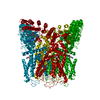
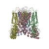
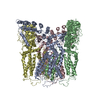
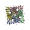
| Title | Structure of Transient Receptor Potential (TRP) channel TRPV6 in the presence of barium | |||||||||
 Components Components | Transient receptor potential cation channel subfamily V member 6 | |||||||||
 Keywords Keywords | TRANSPORT PROTEIN | |||||||||
| Function / homology |  Function and homology information Function and homology informationparathyroid hormone secretion / TRP channels / calcium ion import / calcium-activated cation channel activity / calcium ion import across plasma membrane / calcium ion homeostasis / calcium channel complex / response to calcium ion / calcium ion transmembrane transport / calcium channel activity ...parathyroid hormone secretion / TRP channels / calcium ion import / calcium-activated cation channel activity / calcium ion import across plasma membrane / calcium ion homeostasis / calcium channel complex / response to calcium ion / calcium ion transmembrane transport / calcium channel activity / calcium ion transport / protein homotetramerization / calmodulin binding / apical plasma membrane / metal ion binding / identical protein binding / plasma membrane Similarity search - Function | |||||||||
| Biological species |  | |||||||||
| Method |  X-RAY DIFFRACTION / SYNCHROTRON / MOLECULAR REPLACEMENT / Resolution: 3.85 Å X-RAY DIFFRACTION / SYNCHROTRON / MOLECULAR REPLACEMENT / Resolution: 3.85 Å | |||||||||
 Authors Authors | Saotome, K. / Singh, A.K. / Yelshanskaya, M.V. / Sobolevsky, A.I. | |||||||||
| Funding support |  United States, 2items United States, 2items
| |||||||||
 Citation Citation | Journal: Nature / Year: 2016 Title: Crystal structure of the epithelial calcium channel TRPV6. Authors: Saotome, K. / Singh, A.K. / Yelshanskaya, M.V. / Sobolevsky, A.I. | |||||||||
| History |
|
- Structure visualization
| Structure viewer | Molecule: MolmilJmol/JSmol |
|---|
- Downloads & links
-Download
| PDBx/mmCIF format | 5iwr.cif.gz | 133.4 KB | Display | PDBx/mmCIF format |
|---|---|---|---|---|
| PDB format | pdb5iwr.ent.gz | 102 KB | Display | PDB format |
| PDBx/mmJSON format | 5iwr.json.gz | Tree view | PDBx/mmJSON format | |
| Others |  Other downloads Other downloads |
-Validation report
| Arichive directory | https://data.pdbj.org/pub/pdb/validation_reports/iw/5iwrftp://data.pdbj.org/pub/pdb/validation_reports/iw/5iwr | HTTPS FTP |
|---|
-Related structure data
| Related structure data |  5iwkC  5iwpC  5iwtC  2rfaS C: citing same article ( S: Starting model for refinement |
|---|---|
| Similar structure data |


-Links
 PDBj
PDBj
- Assembly
| Deposited unit | 
| ||||||||||||
|---|---|---|---|---|---|---|---|---|---|---|---|---|---|
| 1 | 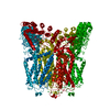
| ||||||||||||
| Unit cell |
| ||||||||||||
| Components on special symmetry positions |
|
-Components
| #1: Protein | Mass: 77148.469 Da / Num. of mol.: 1 / Mutation: I102Y, L132N, M136Q, L535Q Source method: isolated from a genetically manipulated source Source: (gene. exp.)  Homo sapiens (human) / References: UniProt: Q9R186 Homo sapiens (human) / References: UniProt: Q9R186 | ||
|---|---|---|---|
| #2: Chemical | ChemComp-BA /   Mass: 137.327 Da / Num. of mol.: 4 / Source method: obtained synthetically / Formula: Ba Mass: 137.327 Da / Num. of mol.: 4 / Source method: obtained synthetically / Formula: Ba#3: Chemical | ChemComp-DTB / |   Mass: 214.262 Da / Num. of mol.: 1 / Source method: obtained synthetically / Formula: C10H18N2O3 Mass: 214.262 Da / Num. of mol.: 1 / Source method: obtained synthetically / Formula: C10H18N2O3 |
-Experimental details
-Experiment
| Experiment | Method: X-RAY DIFFRACTION / Number of used crystals: 1 |
|---|
- Sample preparation
| Crystal | Density Matthews: 3.85 Å3/Da / Density % sol: 67 % |
|---|---|
| Crystal grow | Temperature: 293 K / Method: vapor diffusion Details: 20-24% PEG 350 MME, 50 mM NaCl, and 50 mM Tris-HCl pH 8.0-8.5 |
-Data collection
| Diffraction | Mean temperature: 100 K |
|---|---|
| Diffraction source | Source: SYNCHROTRON / Site: APS / Beamline: 24-ID-C / Wavelength: 1.75 Å |
| Detector | Type: DECTRIS PILATUS 6M-F / Detector: PIXEL / Date: Mar 24, 2015 |
| Radiation | Protocol: SINGLE WAVELENGTH / Monochromatic (M) / Laue (L): M / Scattering type: x-ray |
| Radiation wavelength | Wavelength: 1.75 Å / Relative weight: 1 |
| Reflection | Resolution: 3.85→49.56 Å / Num. obs: 21705 / % possible obs: 99.5 % / Redundancy: 15.4 % / CC1/2: 0.98 / Rmerge(I) obs: 0.131 / Net I/σ(I): 15.2 |
| Reflection shell | Resolution: 3.85→3.99 Å / Redundancy: 13.9 % / Rmerge(I) obs: 2.285 / Mean I/σ(I) obs: 1.5 / % possible all: 95.4 |
- Processing
| Software |
| |||||||||||||||||||||||||||||||||||||||||||||||||||||||||||||||
|---|---|---|---|---|---|---|---|---|---|---|---|---|---|---|---|---|---|---|---|---|---|---|---|---|---|---|---|---|---|---|---|---|---|---|---|---|---|---|---|---|---|---|---|---|---|---|---|---|---|---|---|---|---|---|---|---|---|---|---|---|---|---|---|---|
| Refinement | Method to determine structure: MOLECULAR REPLACEMENT Starting model: 2RFA Resolution: 3.85→49.556 Å / SU ML: 0.62 / Cross valid method: FREE R-VALUE / σ(F): 1.34 / Phase error: 33.89 / Stereochemistry target values: ML
| |||||||||||||||||||||||||||||||||||||||||||||||||||||||||||||||
| Solvent computation | Shrinkage radii: 0.9 Å / VDW probe radii: 1.11 Å / Solvent model: FLAT BULK SOLVENT MODEL | |||||||||||||||||||||||||||||||||||||||||||||||||||||||||||||||
| Refinement step | Cycle: LAST / Resolution: 3.85→49.556 Å
| |||||||||||||||||||||||||||||||||||||||||||||||||||||||||||||||
| Refine LS restraints |
| |||||||||||||||||||||||||||||||||||||||||||||||||||||||||||||||
| LS refinement shell |
|
