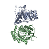
 Movie
Movie Controller


+ Open data

- Basic information
| Entry | Database: PDB / ID: 5ipw | ||||||
|---|---|---|---|---|---|---|---|
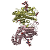
| Title | oligopeptide-binding protein OppA | ||||||
 Components Components | Oligopeptide ABC transporter, periplasmic oligopeptide-binding protein, putative | ||||||
 Keywords Keywords | PEPTIDE BINDING PROTEIN / oligopeptide-binding protein | ||||||
| Function / homology | Solute-binding protein family 5, conserved site / Bacterial extracellular solute-binding proteins, family 5 signature. / Solute-binding protein family 5 domain / Solute-binding protein family 5 / Bacterial extracellular solute-binding proteins, family 5 Middle / peptide transport / peptide transmembrane transporter activity / Oligopeptide ABC transporter, periplasmic oligopeptide-binding protein, putative Function and homology information Function and homology information | ||||||
| Biological species |   Thermotoga maritima MSB8 (bacteria) Thermotoga maritima MSB8 (bacteria) | ||||||
| Method |  X-RAY DIFFRACTION / SYNCHROTRON / MOLECULAR REPLACEMENT / Resolution: 2.6 Å X-RAY DIFFRACTION / SYNCHROTRON / MOLECULAR REPLACEMENT / Resolution: 2.6 Å | ||||||
 Authors Authors | Lee, H.H. / Kim, H.J. / Yoon, H.J. | ||||||
 Citation Citation | Journal: Extremophiles / Year: 2016 Title: Crystal structure of a putative oligopeptide-binding periplasmic protein from a hyperthermophile Authors: Yoon, H.J. / Kim, H.J. / Mikami, B. / Yu, Y.G. / Lee, H.H. | ||||||
| History |
|
- Structure visualization
| Structure viewer | Molecule: MolmilJmol/JSmol |
|---|
- Downloads & links
-Download
| PDBx/mmCIF format | 5ipw.cif.gz | 143.6 KB | Display | PDBx/mmCIF format |
|---|---|---|---|---|
| PDB format | pdb5ipw.ent.gz | 110.9 KB | Display | PDB format |
| PDBx/mmJSON format | 5ipw.json.gz | Tree view | PDBx/mmJSON format | |
| Others |  Other downloads Other downloads |
-Validation report
| Summary document | 5ipw_validation.pdf.gz | 425.1 KB | Display | wwPDB validaton report |
|---|---|---|---|---|
| Full document | 5ipw_full_validation.pdf.gz | 434.4 KB | Display | |
| Data in XML | 5ipw_validation.xml.gz | 25.5 KB | Display | |
| Data in CIF | 5ipw_validation.cif.gz | 35.3 KB | Display | |
| Arichive directory | https://data.pdbj.org/pub/pdb/validation_reports/ip/5ipwftp://data.pdbj.org/pub/pdb/validation_reports/ip/5ipw | HTTPS FTP |
-Related structure data
| Similar structure data |
|---|


-Links
 PDBj
PDBj- Assembly
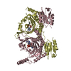
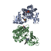
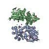
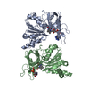
| Deposited unit | 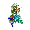
| ||||||||
|---|---|---|---|---|---|---|---|---|---|
| 1 |
| ||||||||
| Unit cell |
|
-Components
| #1: Protein | Mass: 72971.508 Da / Num. of mol.: 1 / Fragment: UNP residues 31-659 Source method: isolated from a genetically manipulated source Source: (gene. exp.) Thermotoga maritima MSB8 (bacteria) / Strain: MSB8 / Gene: TM_0056 / Production host: |
|---|---|
| #2: Water | ChemComp-HOH /  Mass: 18.015 Da / Num. of mol.: 155 / Source method: isolated from a natural source / Formula: H2O Mass: 18.015 Da / Num. of mol.: 155 / Source method: isolated from a natural source / Formula: H2O |
-Experimental details
-Experiment
| Experiment | Method: X-RAY DIFFRACTION / Number of used crystals: 1 |
|---|
- Sample preparation
| Crystal | Density Matthews: 4.14 Å3/Da / Density % sol: 70.3 % |
|---|---|
| Crystal grow | Temperature: 296 K / Method: vapor diffusion, sitting drop / pH: 7.5 / Details: 100 mM sodium HEPES, 1.0 M tri-sodium citrate |
-Data collection
| Diffraction | Mean temperature: 100 K |
|---|---|
| Diffraction source | Source: SYNCHROTRON / Site: SPring-8  / Beamline: BL26B1 / Wavelength: 0.9789 Å / Beamline: BL26B1 / Wavelength: 0.9789 Å |
| Detector | Type: BRUKER SMART 6500 / Detector: CCD / Date: Nov 19, 2012 |
| Radiation | Protocol: SINGLE WAVELENGTH / Monochromatic (M) / Laue (L): M / Scattering type: x-ray |
| Radiation wavelength | Wavelength: 0.9789 Å / Relative weight: 1 |
| Reflection | Resolution: 2.6→50 Å / Num. obs: 37769 / % possible obs: 99.9 % / Redundancy: 13.4 % / Net I/σ(I): 39.2 |
- Processing
| Software |
| ||||||||||||||||||||||||||||||||||||||||||||||||||||||||||||||||||||||||||||||||||||||||||||||||||||||||||||||||||||||||||||||||||||||||||||||||||||||||||||||||||||||||||||||||||||||
|---|---|---|---|---|---|---|---|---|---|---|---|---|---|---|---|---|---|---|---|---|---|---|---|---|---|---|---|---|---|---|---|---|---|---|---|---|---|---|---|---|---|---|---|---|---|---|---|---|---|---|---|---|---|---|---|---|---|---|---|---|---|---|---|---|---|---|---|---|---|---|---|---|---|---|---|---|---|---|---|---|---|---|---|---|---|---|---|---|---|---|---|---|---|---|---|---|---|---|---|---|---|---|---|---|---|---|---|---|---|---|---|---|---|---|---|---|---|---|---|---|---|---|---|---|---|---|---|---|---|---|---|---|---|---|---|---|---|---|---|---|---|---|---|---|---|---|---|---|---|---|---|---|---|---|---|---|---|---|---|---|---|---|---|---|---|---|---|---|---|---|---|---|---|---|---|---|---|---|---|---|---|---|---|
| Refinement | Method to determine structure: MOLECULAR REPLACEMENT / Resolution: 2.6→50 Å / Cor.coef. Fo:Fc: 0.946 / Cor.coef. Fo:Fc free: 0.928 / SU B: 9.49 / SU ML: 0.194 / Cross valid method: THROUGHOUT / ESU R: 0.334 / ESU R Free: 0.244 / Details: HYDROGENS HAVE BEEN ADDED IN THE RIDING POSITIONS
| ||||||||||||||||||||||||||||||||||||||||||||||||||||||||||||||||||||||||||||||||||||||||||||||||||||||||||||||||||||||||||||||||||||||||||||||||||||||||||||||||||||||||||||||||||||||
| Solvent computation | Ion probe radii: 0.8 Å / Shrinkage radii: 0.8 Å / VDW probe radii: 1.2 Å | ||||||||||||||||||||||||||||||||||||||||||||||||||||||||||||||||||||||||||||||||||||||||||||||||||||||||||||||||||||||||||||||||||||||||||||||||||||||||||||||||||||||||||||||||||||||
| Displacement parameters | Biso mean: 60.952 Å2
| ||||||||||||||||||||||||||||||||||||||||||||||||||||||||||||||||||||||||||||||||||||||||||||||||||||||||||||||||||||||||||||||||||||||||||||||||||||||||||||||||||||||||||||||||||||||
| Refinement step | Cycle: LAST / Resolution: 2.6→50 Å
| ||||||||||||||||||||||||||||||||||||||||||||||||||||||||||||||||||||||||||||||||||||||||||||||||||||||||||||||||||||||||||||||||||||||||||||||||||||||||||||||||||||||||||||||||||||||
| Refine LS restraints |
|