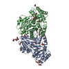
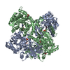
Entry Database : PDB / ID : 5ijgTitle Crystal structure of O-acetylhomoserine sulfhydrolase from Brucella melitensis at 2.0 A resolution Cys/Met metabolism pyridoxal-phosphate-dependent enzyme Keywords / / / / / / Function / homology Function Domain/homology Component
/ / / / / / / / / / / / / / / / / / / / / / / / / / / Biological species Brucella melitensis biotype 1 (bacteria)Method / / / / Resolution : 2 Å Authors Boyko, K.M. / Nikolaeva, A.Y. / Koolikova, V.V. / Kotlov, M.I. / Demidkina, T.V. / Popov, V.O. Journal : To Be Published Title : Crystal structure of O-acetylhomoserine sulfhydrolase from Brucella melitensis at 2.0 A resolutionAuthors : Boyko, K.M. / Nikolaeva, A.Y. / Koolikova, V. / Demidkina, T.V. / Popov, V.O. History Deposition Mar 2, 2016 Deposition site / Processing site Revision 1.0 Apr 5, 2017 Provider / Type Revision 1.1 Jan 10, 2024 Group / Database references / Refinement descriptionCategory chem_comp_atom / chem_comp_bond ... chem_comp_atom / chem_comp_bond / database_2 / pdbx_initial_refinement_model Item / _database_2.pdbx_database_accession
Show all Show less
 Movie
Movie Controller

 Yorodumi
 Open data
 Basic information
 Components
 Keywords
 Function and homology information
 Brucella melitensis biotype 1 (bacteria)
 X-RAY DIFFRACTION /
  Authors
 Citation
 Structure visualization
 Downloads & links
 Other downloads

 PDBj
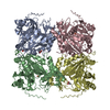
PDBj Assembly
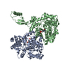

 Mass: 247.142 Da / Num. of mol.: 2 / Source method: obtained synthetically / Formula: C8H10NO6P
 Mass: 92.094 Da / Num. of mol.: 1 / Source method: obtained synthetically / Formula: C3H8O3
 Mass: 18.015 Da / Num. of mol.: 487 / Source method: isolated from a natural source / Formula: H2O
 Sample preparation
 / Beamline: BL41XU / Wavelength: 1 Å
 Processing