 Movie
Movie Controller

[English] 日本語
 Yorodumi
- PDB-5iet: Crystal Structure of Mycobacterium Tuberculosis ATP-independent P... -
+ Open data

- Basic information
| Entry | Database: PDB / ID: 5iet | ||||||
|---|---|---|---|---|---|---|---|
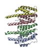
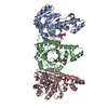
| Title | Crystal Structure of Mycobacterium Tuberculosis ATP-independent Proteasome activator | ||||||
 Components Components | Bacterial proteasome activator | ||||||
 Keywords Keywords | GENE REGULATION / Activator / Apoptosis | ||||||
| Function / homology |  Function and homology information Function and homology informationproteasome accessory complex / positive regulation of proteasomal protein catabolic process / proteasome binding / peptidoglycan-based cell wall / protein homooligomerization / plasma membrane Similarity search - Function | ||||||
| Biological species |   Mycobacterium tuberculosis (bacteria) Mycobacterium tuberculosis (bacteria) | ||||||
| Method |  X-RAY DIFFRACTION / SYNCHROTRON / SAD / Resolution: 2.88 Å X-RAY DIFFRACTION / SYNCHROTRON / SAD / Resolution: 2.88 Å | ||||||
 Authors Authors | Bai, L. / Hu, K. / Wang, T. / Jastrab, J.B. / Darwin, K.H. / Li, H. | ||||||
| Funding support |  United States, 1items United States, 1items
| ||||||
 Citation Citation | Journal: Proc.Natl.Acad.Sci.USA / Year: 2016 Title: Structural analysis of the dodecameric proteasome activator PafE in Mycobacterium tuberculosis. Authors: Bai, L. / Hu, K. / Wang, T. / Jastrab, J.B. / Darwin, K.H. / Li, H. | ||||||
| History |
|
- Structure visualization
| Structure viewer | Molecule: MolmilJmol/JSmol |
|---|
- Downloads & links
-Download
| PDBx/mmCIF format | 5iet.cif.gz | 57.2 KB | Display | PDBx/mmCIF format |
|---|---|---|---|---|
| PDB format | pdb5iet.ent.gz | 44.4 KB | Display | PDB format |
| PDBx/mmJSON format | 5iet.json.gz | Tree view | PDBx/mmJSON format | |
| Others |  Other downloads Other downloads |
-Validation report
| Summary document | 5iet_validation.pdf.gz | 461.7 KB | Display | wwPDB validaton report |
|---|---|---|---|---|
| Full document | 5iet_full_validation.pdf.gz | 461.9 KB | Display | |
| Data in XML | 5iet_validation.xml.gz | 11 KB | Display | |
| Data in CIF | 5iet_validation.cif.gz | 14.5 KB | Display | |
| Arichive directory | https://data.pdbj.org/pub/pdb/validation_reports/ie/5ietftp://data.pdbj.org/pub/pdb/validation_reports/ie/5iet | HTTPS FTP |
-Related structure data


-Links
 PDBj
PDBj- Assembly
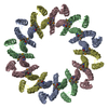
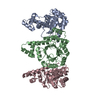
| Deposited unit | 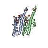
| ||||||||
|---|---|---|---|---|---|---|---|---|---|
| 1 | x 6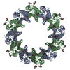
| ||||||||
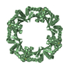
| Unit cell |
|
-Components
| #1: Protein | Mass: 17133.516 Da / Num. of mol.: 2 / Fragment: residues 19-157 Source method: isolated from a genetically manipulated source Source: (gene. exp.) Mycobacterium tuberculosis (bacteria) / Gene: AFL40_3934 / Production host: #2: Chemical | ChemComp-SO4 /   Mass: 96.063 Da / Num. of mol.: 4 / Source method: obtained synthetically / Formula: SO4 Mass: 96.063 Da / Num. of mol.: 4 / Source method: obtained synthetically / Formula: SO4#3: Chemical | ChemComp-MPD / (   Mass: 118.174 Da / Num. of mol.: 4 / Source method: obtained synthetically / Formula: C6H14O2 / Comment: precipitant*YM Mass: 118.174 Da / Num. of mol.: 4 / Source method: obtained synthetically / Formula: C6H14O2 / Comment: precipitant*YM#4: Water | ChemComp-HOH / |  Mass: 18.015 Da / Num. of mol.: 74 / Source method: isolated from a natural source / Formula: H2O Mass: 18.015 Da / Num. of mol.: 74 / Source method: isolated from a natural source / Formula: H2O |
|---|
-Experimental details
-Experiment
| Experiment | Method: X-RAY DIFFRACTION / Number of used crystals: 1 |
|---|
- Sample preparation
| Crystal | Density Matthews: 4.89 Å3/Da / Density % sol: 74.85 % |
|---|---|
| Crystal grow | Temperature: 293 K / Method: vapor diffusion, sitting drop / pH: 5.9 Details: 0.2 M ammonium acetate, 0.1 M sodium citrate 29% 2-methyl-2,4-pentanediol |
-Data collection
| Diffraction | Mean temperature: 100 K |
|---|---|
| Diffraction source | Source: SYNCHROTRON / Site: APS / Beamline: 31-ID / Wavelength: 0.9793 Å |
| Detector | Type: RAYONIX MX-225 / Detector: CCD / Date: Nov 25, 2014 |
| Radiation | Protocol: SINGLE WAVELENGTH / Monochromatic (M) / Laue (L): M / Scattering type: x-ray |
| Radiation wavelength | Wavelength: 0.9793 Å / Relative weight: 1 |
| Reflection | Resolution: 2.88→42 Å / Num. obs: 16296 / % possible obs: 99.9 % / Redundancy: 19.3 % / Net I/σ(I): 15.7 |
- Processing
| Software |
| ||||||||||||||||||||||||||||||||||||||||||||||||||||||||||||||||||||||||||||||||||||||||||||||||||||||||||||||||||||||||||||||||||||||||||||||||||||||||||||||||||||||||||||||||||||||
|---|---|---|---|---|---|---|---|---|---|---|---|---|---|---|---|---|---|---|---|---|---|---|---|---|---|---|---|---|---|---|---|---|---|---|---|---|---|---|---|---|---|---|---|---|---|---|---|---|---|---|---|---|---|---|---|---|---|---|---|---|---|---|---|---|---|---|---|---|---|---|---|---|---|---|---|---|---|---|---|---|---|---|---|---|---|---|---|---|---|---|---|---|---|---|---|---|---|---|---|---|---|---|---|---|---|---|---|---|---|---|---|---|---|---|---|---|---|---|---|---|---|---|---|---|---|---|---|---|---|---|---|---|---|---|---|---|---|---|---|---|---|---|---|---|---|---|---|---|---|---|---|---|---|---|---|---|---|---|---|---|---|---|---|---|---|---|---|---|---|---|---|---|---|---|---|---|---|---|---|---|---|---|---|
| Refinement | Method to determine structure: SAD / Resolution: 2.88→41.99 Å / Cor.coef. Fo:Fc: 0.923 / Cor.coef. Fo:Fc free: 0.901 / SU B: 6.806 / SU ML: 0.134 / Cross valid method: THROUGHOUT / ESU R: 0.278 / ESU R Free: 0.219 / Details: HYDROGENS HAVE BEEN ADDED IN THE RIDING POSITIONS
| ||||||||||||||||||||||||||||||||||||||||||||||||||||||||||||||||||||||||||||||||||||||||||||||||||||||||||||||||||||||||||||||||||||||||||||||||||||||||||||||||||||||||||||||||||||||
| Solvent computation | Ion probe radii: 0.8 Å / Shrinkage radii: 0.8 Å / VDW probe radii: 1.2 Å | ||||||||||||||||||||||||||||||||||||||||||||||||||||||||||||||||||||||||||||||||||||||||||||||||||||||||||||||||||||||||||||||||||||||||||||||||||||||||||||||||||||||||||||||||||||||
| Displacement parameters | Biso mean: 30.634 Å2
| ||||||||||||||||||||||||||||||||||||||||||||||||||||||||||||||||||||||||||||||||||||||||||||||||||||||||||||||||||||||||||||||||||||||||||||||||||||||||||||||||||||||||||||||||||||||
| Refinement step | Cycle: 1 / Resolution: 2.88→41.99 Å
| ||||||||||||||||||||||||||||||||||||||||||||||||||||||||||||||||||||||||||||||||||||||||||||||||||||||||||||||||||||||||||||||||||||||||||||||||||||||||||||||||||||||||||||||||||||||
| Refine LS restraints |
|