 Movie
Movie Controller

[English] 日本語
 Yorodumi
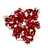
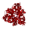
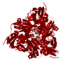
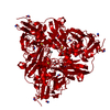
- PDB-5i6k: Crystal Structure of Copper Nitrite Reductase at 100K after 0.69 MGy -
+ Open data

- Basic information
| Entry | Database: PDB / ID: 5i6k | |||||||||
|---|---|---|---|---|---|---|---|---|---|---|
| Title | Crystal Structure of Copper Nitrite Reductase at 100K after 0.69 MGy | |||||||||
 Components Components | Copper-containing nitrite reductase | |||||||||
 Keywords Keywords | OXIDOREDUCTASE / Copper Nitrite Reductase / Reaction Mechanism / Serial Crystallography | |||||||||
| Function / homology |  Function and homology information Function and homology informationdenitrification pathway / nitrite reductase (NO-forming) / nitrite reductase (NO-forming) activity / nitrate assimilation / periplasmic space / copper ion binding Similarity search - Function | |||||||||
| Biological species |  Achromobacter cycloclastes (bacteria) Achromobacter cycloclastes (bacteria) | |||||||||
| Method |  X-RAY DIFFRACTION / SYNCHROTRON / MOLECULAR REPLACEMENT / Resolution: 1.07 Å X-RAY DIFFRACTION / SYNCHROTRON / MOLECULAR REPLACEMENT / Resolution: 1.07 Å | |||||||||
 Authors Authors | Horrell, S. / Hough, M.A. / Strange, R.W. | |||||||||
| Funding support |  United Kingdom, 2items United Kingdom, 2items
| |||||||||
 Citation Citation | Journal: Iucrj / Year: 2016 Title: Serial crystallography captures enzyme catalysis in copper nitrite reductase at atomic resolution from one crystal. Authors: Horrell, S. / Antonyuk, S.V. / Eady, R.R. / Hasnain, S.S. / Hough, M.A. / Strange, R.W. | |||||||||
| History |
|
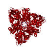
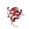
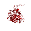
- Structure visualization
| Structure viewer | Molecule: MolmilJmol/JSmol |
|---|
- Downloads & links
-Download
| PDBx/mmCIF format | 5i6k.cif.gz | 166.2 KB | Display | PDBx/mmCIF format |
|---|---|---|---|---|
| PDB format | pdb5i6k.ent.gz | 129.9 KB | Display | PDB format |
| PDBx/mmJSON format | 5i6k.json.gz | Tree view | PDBx/mmJSON format | |
| Others |  Other downloads Other downloads |
-Validation report
| Arichive directory | https://data.pdbj.org/pub/pdb/validation_reports/i6/5i6kftp://data.pdbj.org/pub/pdb/validation_reports/i6/5i6k | HTTPS FTP |
|---|
-Related structure data
| Related structure data | 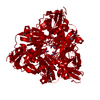 5i6lC  5i6mC  5i6nC  5i6oC  5i6pC  2bwiS S: Starting model for refinement C: citing same article ( |
|---|---|
| Similar structure data |


-Links
 PDBj
PDBj

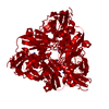
- Assembly
| Deposited unit | 
| ||||||||
|---|---|---|---|---|---|---|---|---|---|
| 1 | 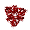
| ||||||||
| Unit cell |
|
-Components
| #1: Protein | Mass: 36621.332 Da / Num. of mol.: 1 / Fragment: Copper Nitrite Reductase Source method: isolated from a genetically manipulated source Source: (gene. exp.) Achromobacter cycloclastes (bacteria) / Gene: nirK / Plasmid: pET-26b(+) / Production host: | ||||
|---|---|---|---|---|---|
| #2: Chemical |   Mass: 63.546 Da / Num. of mol.: 2 / Source method: obtained synthetically / Formula: Cu Mass: 63.546 Da / Num. of mol.: 2 / Source method: obtained synthetically / Formula: Cu#3: Chemical | ChemComp-NO2 / 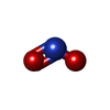  Mass: 46.005 Da / Num. of mol.: 6 / Source method: obtained synthetically / Formula: NO2 Mass: 46.005 Da / Num. of mol.: 6 / Source method: obtained synthetically / Formula: NO2#4: Water | ChemComp-HOH / |  Mass: 18.015 Da / Num. of mol.: 360 / Source method: isolated from a natural source / Formula: H2O Mass: 18.015 Da / Num. of mol.: 360 / Source method: isolated from a natural source / Formula: H2O |
-Experimental details
-Experiment
| Experiment | Method: X-RAY DIFFRACTION / Number of used crystals: 1 |
|---|
- Sample preparation
| Crystal | Density Matthews: 1.98 Å3/Da / Density % sol: 37.91 % / Mosaicity: 0.9 ° |
|---|---|
| Crystal grow | Temperature: 298 K / Method: evaporation / pH: 4.5 Details: 1.7 M Ammonium Sulphate, 0.1 M Sodium Acetate pH 4.5 |
-Data collection
| Diffraction | Mean temperature: 100 K | ||||||||||||||||||||||||||||||
|---|---|---|---|---|---|---|---|---|---|---|---|---|---|---|---|---|---|---|---|---|---|---|---|---|---|---|---|---|---|---|---|
| Diffraction source | Source: SYNCHROTRON / Site: Diamond / Beamline: I04 / Wavelength: 0.97 Å | ||||||||||||||||||||||||||||||
| Detector | Type: DECTRIS PILATUS3 6M / Detector: PIXEL / Date: Dec 14, 2013 | ||||||||||||||||||||||||||||||
| Radiation | Protocol: SINGLE WAVELENGTH / Monochromatic (M) / Laue (L): M / Scattering type: x-ray | ||||||||||||||||||||||||||||||
| Radiation wavelength | Wavelength: 0.97 Å / Relative weight: 1 | ||||||||||||||||||||||||||||||
| Reflection | Resolution: 1.07→42.61 Å / Num. obs: 121734 / % possible obs: 96.7 % / Redundancy: 3.9 % / CC1/2: 0.999 / Rmerge(I) obs: 0.048 / Rpim(I) all: 0.026 / Rrim(I) all: 0.054 / Net I/σ(I): 13.4 / Num. measured all: 475646 / Scaling rejects: 1 | ||||||||||||||||||||||||||||||
| Reflection shell | Diffraction-ID: 1 / Rejects: _
|
- Processing
| Software |
| ||||||||||||||||||||||||||||||||||||||||||||||||||||||||||||||||||||||||||||||||||||||||||
|---|---|---|---|---|---|---|---|---|---|---|---|---|---|---|---|---|---|---|---|---|---|---|---|---|---|---|---|---|---|---|---|---|---|---|---|---|---|---|---|---|---|---|---|---|---|---|---|---|---|---|---|---|---|---|---|---|---|---|---|---|---|---|---|---|---|---|---|---|---|---|---|---|---|---|---|---|---|---|---|---|---|---|---|---|---|---|---|---|---|---|---|
| Refinement | Method to determine structure: MOLECULAR REPLACEMENT Starting model: 2BWI Resolution: 1.07→42.61 Å / Cor.coef. Fo:Fc: 0.972 / Cor.coef. Fo:Fc free: 0.967 / SU B: 0.953 / SU ML: 0.02 / Cross valid method: THROUGHOUT / σ(F): 0 / ESU R: 0.029 / ESU R Free: 0.03 / Stereochemistry target values: MAXIMUM LIKELIHOOD Details: HYDROGENS HAVE BEEN ADDED IN THE RIDING POSITIONS U VALUES : REFINED INDIVIDUALLY
| ||||||||||||||||||||||||||||||||||||||||||||||||||||||||||||||||||||||||||||||||||||||||||
| Solvent computation | Ion probe radii: 0.8 Å / Shrinkage radii: 0.8 Å / VDW probe radii: 1.2 Å / Solvent model: MASK | ||||||||||||||||||||||||||||||||||||||||||||||||||||||||||||||||||||||||||||||||||||||||||
| Displacement parameters | Biso max: 91.22 Å2 / Biso mean: 14.026 Å2 / Biso min: 6.96 Å2
| ||||||||||||||||||||||||||||||||||||||||||||||||||||||||||||||||||||||||||||||||||||||||||
| Refinement step | Cycle: final / Resolution: 1.07→42.61 Å
| ||||||||||||||||||||||||||||||||||||||||||||||||||||||||||||||||||||||||||||||||||||||||||
| Refine LS restraints |
| ||||||||||||||||||||||||||||||||||||||||||||||||||||||||||||||||||||||||||||||||||||||||||
| LS refinement shell | Resolution: 1.07→1.098 Å / Total num. of bins used: 20
|
