[English] 日本語
Yorodumi
- PDB-5hvz: Crystal structure of smAKAP AKB domain bound RIa dimerization/doc... -

+
Open data


ID or keywords:

Loading...

-
Basic information

Entry
Database: PDB / ID: 5hvz
TitleCrystal structure of smAKAP AKB domain bound RIa dimerization/docking (D/D) complex at 2.0 A resolution
Components
  • Small membrane A-kinase anchor protein
  • cAMP-dependent protein kinase type I-alpha regulatory subunit
KeywordsTRANSFERASE / A-kinase anchoring protein / small membrane AKAP / protein kinase A / regulatory subunit
Function / homology
Function and homology information


: / PKA activation in glucagon signalling / DARPP-32 events / CREB1 phosphorylation through the activation of Adenylate Cyclase / GPER1 signaling / Factors involved in megakaryocyte development and platelet production / PKA activation / nucleotide-activated protein kinase complex / Hedgehog 'off' state / cAMP-dependent protein kinase inhibitor activity ...: / PKA activation in glucagon signalling / DARPP-32 events / CREB1 phosphorylation through the activation of Adenylate Cyclase / GPER1 signaling / Factors involved in megakaryocyte development and platelet production / PKA activation / nucleotide-activated protein kinase complex / Hedgehog 'off' state / cAMP-dependent protein kinase inhibitor activity / cardiac muscle cell proliferation / cAMP-dependent protein kinase complex / sarcomere organization / Vasopressin regulates renal water homeostasis via Aquaporins / cellular response to glucagon stimulus / protein kinase A regulatory subunit binding / negative regulation of activated T cell proliferation / plasma membrane raft / protein kinase A catalytic subunit binding / axoneme / mesoderm formation / immunological synapse / cAMP binding / multivesicular body / regulation of protein phosphorylation / neuromuscular junction / positive regulation of insulin secretion / adenylate cyclase-activating G protein-coupled receptor signaling pathway / protein domain specific binding / negative regulation of gene expression / centrosome / glutamatergic synapse / ubiquitin protein ligase binding / identical protein binding / plasma membrane / cytosol / cytoplasm
Similarity search - Function
Small membrane A-kinase anchor protein / Small membrane A-kinase anchor protein / cAMP-dependent protein kinase regulatory subunit, dimerization-anchoring domain / cAMP-dependent Protein Kinase, Chain A / : / cAMP-dependent protein kinase regulatory subunit / cAMP-dependent protein kinase regulatory subunit, dimerization-anchoring domain / Regulatory subunit of type II PKA R-subunit / RIIalpha, Regulatory subunit portion of type II PKA R-subunit / Cyclic nucleotide-binding domain signature 2. ...Small membrane A-kinase anchor protein / Small membrane A-kinase anchor protein / cAMP-dependent protein kinase regulatory subunit, dimerization-anchoring domain / cAMP-dependent Protein Kinase, Chain A / : / cAMP-dependent protein kinase regulatory subunit / cAMP-dependent protein kinase regulatory subunit, dimerization-anchoring domain / Regulatory subunit of type II PKA R-subunit / RIIalpha, Regulatory subunit portion of type II PKA R-subunit / Cyclic nucleotide-binding domain signature 2. / Cyclic nucleotide-binding domain signature 1. / Cyclic nucleotide-binding, conserved site / Cyclic nucleotide-monophosphate binding domain / Cyclic nucleotide-binding domain / cAMP/cGMP binding motif profile. / Cyclic nucleotide-binding domain / Cyclic nucleotide-binding domain superfamily / RmlC-like jelly roll fold / Up-down Bundle / Mainly Alpha
Similarity search - Domain/homology
cAMP-dependent protein kinase type I-alpha regulatory subunit / Small membrane A-kinase anchor protein
Similarity search - Component
Biological speciesBos taurus (cattle)
Homo sapiens (human)
MethodX-RAY DIFFRACTION / SYNCHROTRON / MOLECULAR REPLACEMENT / Resolution: 2 Å
AuthorsWu, J. / Burgers, P.P. / Bruystens, J. / Heck, A.J.R. / Taylor, S.S.
Funding support Netherlands, United States, 2items
OrganizationGrant numberCountry
Netherlands Organization for Scientific Research262067 Netherlands
National Institutes of Health/National Institute of Diabetes and Digestive and Kidney Disease (NIH/NIDDK)DK54441 United States
CitationJournal: Febs J. / Year: 2016
Title: Structure of smAKAP and its regulation by PKA-mediated phosphorylation.
Authors: Burgers, P.P. / Bruystens, J. / Burnley, R.J. / Nikolaev, V.O. / Keshwani, M. / Wu, J. / Janssen, B.J. / Taylor, S.S. / Heck, A.J. / Scholten, A.
History
DepositionJan 28, 2016Deposition site: RCSB / Processing site: RCSB
Revision 1.0Apr 20, 2016Provider: repository / Type: Initial release
Revision 1.1Jun 15, 2016Group: Database references
Revision 1.2Sep 6, 2017Group: Author supporting evidence / Derived calculations / Category: pdbx_audit_support / pdbx_struct_oper_list
Item: _pdbx_audit_support.funding_organization / _pdbx_struct_oper_list.symmetry_operation
Revision 1.3Dec 25, 2019Group: Author supporting evidence / Category: pdbx_audit_support / Item: _pdbx_audit_support.funding_organization
Revision 1.4Sep 27, 2023Group: Data collection / Database references / Refinement description
Category: chem_comp_atom / chem_comp_bond ...chem_comp_atom / chem_comp_bond / database_2 / pdbx_initial_refinement_model
Item: _database_2.pdbx_DOI / _database_2.pdbx_database_accession

-
Structure visualization

Structure viewerMolecule:
MolmilJmol/JSmol

Downloads & links

-
Assembly

Deposited unit
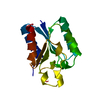
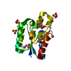
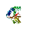
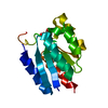
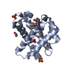
A: cAMP-dependent protein kinase type I-alpha regulatory subunit
B: cAMP-dependent protein kinase type I-alpha regulatory subunit
C: Small membrane A-kinase anchor protein


Theoretical massNumber of molelcules
Total (without water)14,8043
Polymers14,8043
Non-polymers00
Water52229
1


  • Idetical with deposited unit
  • defined by author&software
TypeNameSymmetry operationNumber
identity operation1_555x,y,z1
Buried area4260 Å2
ΔGint-53 kcal/mol
Surface area7000 Å2
MethodPISA
Unit cell
Length a, b, c (Å)37.511, 55.737, 57.268
Angle α, β, γ (deg.)90.00, 90.00, 90.00
Int Tables number19
Space group name H-MP212121

-
Components

#1: Protein/peptide cAMP-dependent protein kinase type I-alpha regulatory subunit


Mass: 6005.980 Da / Num. of mol.: 2
Source method: isolated from a genetically manipulated source
Source: (gene. exp.) Bos taurus (cattle) / Gene: PRKAR1A / Cell line (production host): BL21 / Production host: Escherichia coli (E. coli) / References: UniProt: P00514
#2: Protein/peptide Small membrane A-kinase anchor protein / smAKAP


Mass: 2792.194 Da / Num. of mol.: 1 / Fragment: UNP Residues 56-79 / Source method: obtained synthetically / Source: (synth.) Homo sapiens (human) / References: UniProt: Q9BSF0
#3: Water ChemComp-HOH / water


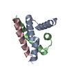
Mass: 18.015 Da / Num. of mol.: 29 / Source method: isolated from a natural source / Formula: H2O

-
Experimental details

-
Experiment

ExperimentMethod: X-RAY DIFFRACTION / Number of used crystals: 1

-
Sample preparation

CrystalDensity Matthews: 2.02 Å3/Da / Density % sol: 39.17 %
Crystal growTemperature: 297 K / Method: microbatch / pH: 3.5
Details: a 2:3 ratio of protein solution:crystallizing (crystallizing solution: 0.1 M Citric acid pH 3.5, 28% w/v Polyethylene glycol 8,000)
Temp details: room temperature

-
Data collection

DiffractionMean temperature: 200 K / Ambient temp details: cryo under liquid nitrogen
Diffraction sourceSource: SYNCHROTRON / Site: ALS / Beamline: 8.2.2 / Wavelength: 1 Å
DetectorType: ADSC QUANTUM 315 / Detector: CCD / Date: Mar 14, 2012
RadiationProtocol: SINGLE WAVELENGTH / Monochromatic (M) / Laue (L): M / Scattering type: x-ray
Radiation wavelengthWavelength: 1 Å / Relative weight: 1
ReflectionResolution: 2→50 Å / Num. obs: 8480 / % possible obs: 98 % / Redundancy: 5.2 % / Rmerge(I) obs: 0.076 / Rsym value: 0.061 / Net I/σ(I): 26.9
Reflection shellResolution: 2→2.05 Å / Redundancy: 5.4 % / Rmerge(I) obs: 0.46 / Mean I/σ(I) obs: 4 / % possible all: 100

-
Processing

Software
NameVersionClassification
REFMAC5.5.0109refinement
HKL-2000data reduction
SCALEPACKdata scaling
PHASERphasing
RefinementMethod to determine structure: MOLECULAR REPLACEMENT
Starting model: 3IM4
Resolution: 2→39.94 Å / Cor.coef. Fo:Fc: 0.939 / Cor.coef. Fo:Fc free: 0.917 / SU B: 3.823 / SU ML: 0.111 / Cross valid method: THROUGHOUT / ESU R: 0.214 / ESU R Free: 0.175 / Details: HYDROGENS HAVE BEEN ADDED IN THE RIDING POSITIONS
RfactorNum. reflection% reflectionSelection details
Rfree0.24133 392 4.7 %RANDOM
Rwork0.20976 ---
obs0.21127 7937 97.53 %-
Solvent computationIon probe radii: 0.8 Å / Shrinkage radii: 0.8 Å / VDW probe radii: 1.4 Å
Displacement parametersBiso mean: 30.44 Å2
Baniso -1Baniso -2Baniso -3
1--0.43 Å20 Å20 Å2
2--0.65 Å20 Å2
3----0.22 Å2
Refinement stepCycle: 1 / Resolution: 2→39.94 Å
ProteinNucleic acidLigandSolventTotal
Num. atoms959 0 0 29 988
Refine LS restraints
Refine-IDTypeDev idealDev ideal targetNumber
X-RAY DIFFRACTIONr_bond_refined_d0.0230.022974
X-RAY DIFFRACTIONr_bond_other_d0.024
X-RAY DIFFRACTIONr_angle_refined_deg1.8911.9881313
X-RAY DIFFRACTIONr_angle_other_deg
X-RAY DIFFRACTIONr_dihedral_angle_1_deg5.4395117
X-RAY DIFFRACTIONr_dihedral_angle_2_deg37.18424.34846
X-RAY DIFFRACTIONr_dihedral_angle_3_deg18.68215182
X-RAY DIFFRACTIONr_dihedral_angle_4_deg23.253157
X-RAY DIFFRACTIONr_chiral_restr0.1510.2151
X-RAY DIFFRACTIONr_gen_planes_refined0.0090.021717
X-RAY DIFFRACTIONr_gen_planes_other
X-RAY DIFFRACTIONr_nbd_refined
X-RAY DIFFRACTIONr_nbd_other
X-RAY DIFFRACTIONr_nbtor_refined
X-RAY DIFFRACTIONr_nbtor_other
X-RAY DIFFRACTIONr_xyhbond_nbd_refined
X-RAY DIFFRACTIONr_xyhbond_nbd_other
X-RAY DIFFRACTIONr_metal_ion_refined
X-RAY DIFFRACTIONr_metal_ion_other
X-RAY DIFFRACTIONr_symmetry_vdw_refined
X-RAY DIFFRACTIONr_symmetry_vdw_other
X-RAY DIFFRACTIONr_symmetry_hbond_refined
X-RAY DIFFRACTIONr_symmetry_hbond_other
X-RAY DIFFRACTIONr_symmetry_metal_ion_refined
X-RAY DIFFRACTIONr_symmetry_metal_ion_other
X-RAY DIFFRACTIONr_mcbond_it1.4371.5602
X-RAY DIFFRACTIONr_mcbond_other
X-RAY DIFFRACTIONr_mcangle_it2.6562959
X-RAY DIFFRACTIONr_mcangle_other
X-RAY DIFFRACTIONr_scbond_it4.3453372
X-RAY DIFFRACTIONr_scbond_other
X-RAY DIFFRACTIONr_scangle_it7.2584.5354
X-RAY DIFFRACTIONr_scangle_other
X-RAY DIFFRACTIONr_long_range_B_refined
X-RAY DIFFRACTIONr_long_range_B_other
X-RAY DIFFRACTIONr_rigid_bond_restr
X-RAY DIFFRACTIONr_sphericity_free
X-RAY DIFFRACTIONr_sphericity_bonded
LS refinement shellResolution: 2→2.052 Å / Total num. of bins used: 20
RfactorNum. reflection% reflection
Rfree0.19 29 -
Rwork0.206 587 -
obs--99.84 %

+
About Yorodumi

-
News

-
Feb 9, 2022. New format data for meta-information of EMDB entries

New format data for meta-information of EMDB entries

  • Version 3 of the EMDB header file is now the official format.
  • The previous official version 1.9 will be removed from the archive.

Related info.:EMDB header

External links:wwPDB to switch to version 3 of the EMDB data model

-
Aug 12, 2020. Covid-19 info

Covid-19 info

URL: https://pdbjlc1.pdbj.org/emnavi/covid19.php

New page: Covid-19 featured information page in EM Navigator.

Related info.:Covid-19 info / Mar 5, 2020. Novel coronavirus structure data

+
Mar 5, 2020. Novel coronavirus structure data

Novel coronavirus structure data

Related info.:Yorodumi Speices / Aug 12, 2020. Covid-19 info

External links:COVID-19 featured content - PDBj / Molecule of the Month (242):Coronavirus Proteases

+
Jan 31, 2019. EMDB accession codes are about to change! (news from PDBe EMDB page)

EMDB accession codes are about to change! (news from PDBe EMDB page)

  • The allocation of 4 digits for EMDB accession codes will soon come to an end. Whilst these codes will remain in use, new EMDB accession codes will include an additional digit and will expand incrementally as the available range of codes is exhausted. The current 4-digit format prefixed with “EMD-” (i.e. EMD-XXXX) will advance to a 5-digit format (i.e. EMD-XXXXX), and so on. It is currently estimated that the 4-digit codes will be depleted around Spring 2019, at which point the 5-digit format will come into force.
  • The EM Navigator/Yorodumi systems omit the EMD- prefix.

Related info.:Q: What is EMD? / ID/Accession-code notation in Yorodumi/EM Navigator

External links:EMDB Accession Codes are Changing Soon! / Contact to PDBj

+
Jul 12, 2017. Major update of PDB

Major update of PDB

  • wwPDB released updated PDB data conforming to the new PDBx/mmCIF dictionary.
  • This is a major update changing the version number from 4 to 5, and with Remediation, in which all the entries are updated.
  • In this update, many items about electron microscopy experimental information are reorganized (e.g. em_software).
  • Now, EM Navigator and Yorodumi are based on the updated data.

External links:wwPDB Remediation / Enriched Model Files Conforming to OneDep Data Standards Now Available in the PDB FTP Archive

-
Yorodumi

Thousand views of thousand structures

  • Yorodumi is a browser for structure data from EMDB, PDB, SASBDB, etc.
  • This page is also the successor to EM Navigator detail page, and also detail information page/front-end page for Omokage search.
  • The word "yorodu" (or yorozu) is an old Japanese word meaning "ten thousand". "mi" (miru) is to see.

Related info.:EMDB / PDB / SASBDB / Comparison of 3 databanks / Yorodumi Search / Aug 31, 2016. New EM Navigator & Yorodumi / Yorodumi Papers / Jmol/JSmol / Function and homology information / Changes in new EM Navigator and Yorodumi

Read more