[English] 日本語
Yorodumi
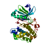
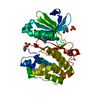
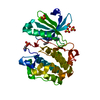
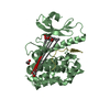
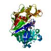
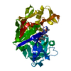
- PDB-5hko: Crystal structure of ABC transporter Solute Binding Protein MSMEG... -

+
Open data


ID or keywords:

Loading...

-
Basic information

Entry
Database: PDB / ID: 5hko
TitleCrystal structure of ABC transporter Solute Binding Protein MSMEG_3598 from Mycobacterium smegmatis str. MC2 155, target EFI-510969, in complex with L-sorbitol
ComponentsABC transporter, carbohydrate uptake transporter-2 (CUT2) family, periplasmic sugar-binding protein
KeywordsSOLUTE-BINDING PROTEIN / ABC TRANSPORTER SOLUTE BINDING PROTEIN / ENZYME FUNCTION INITIATIVE / EFI / Structural Genomics
Function / homology
Function and homology information


cellular response to carbohydrate stimulus / carbohydrate transport / carbohydrate binding / plasma membrane
Similarity search - Function
Periplasmic binding protein / Periplasmic binding protein domain / Response regulator / Periplasmic binding protein-like I / Prokaryotic membrane lipoprotein lipid attachment site profile. / Rossmann fold / 3-Layer(aba) Sandwich / Alpha Beta
Similarity search - Domain/homology
L-glucitol / ACETATE ION / IMIDAZOLE / Xylitol-binding protein
Similarity search - Component
Biological speciesMycobacterium smegmatis (bacteria)
MethodX-RAY DIFFRACTION / SYNCHROTRON / MOLECULAR REPLACEMENT / Resolution: 1.2 Å
AuthorsRoth, Y. / Vetting, M.W. / Al Obaidi, N.F. / Toro, R. / Morisco, L.L. / Benach, J. / Koss, J. / Wasserman, S.R. / Gerlt, J.A. / Almo, S.C. / Enzyme Function Initiative (EFI)
Funding support United States, 1items
OrganizationGrant numberCountry
National Institutes of Health/National Institute of General Medical Sciences (NIH/NIGMS)GM093342 United States
CitationJournal: To be published
Title: Crystal structure of ABC transporter Solute Binding Protein MSMEG_3598 from Mycobacterium smegmatis str. MC2 155, target EFI-510969, in complex with L-sorbitol
Authors: Roth, Y. / Vetting, M.W. / Al Obaidi, N.F. / Toro, R. / Morisco, L.L. / Benach, J. / Koss, J. / Wasserman, S.R. / Gerlt, J.A. / Almo, S.C. / Enzyme Function Initiative (EFI)
History
DepositionJan 14, 2016Deposition site: RCSB / Processing site: RCSB
Revision 1.0Feb 3, 2016Provider: repository / Type: Initial release
Revision 1.1Sep 27, 2017Group: Author supporting evidence / Derived calculations / Category: pdbx_audit_support / pdbx_struct_oper_list
Item: _pdbx_audit_support.funding_organization / _pdbx_struct_oper_list.symmetry_operation
Revision 1.2Dec 25, 2019Group: Author supporting evidence / Category: pdbx_audit_support / Item: _pdbx_audit_support.funding_organization
Revision 1.3Sep 27, 2023Group: Data collection / Database references ...Data collection / Database references / Derived calculations / Refinement description
Category: chem_comp_atom / chem_comp_bond ...chem_comp_atom / chem_comp_bond / database_2 / pdbx_initial_refinement_model / pdbx_struct_conn_angle / struct_conn
Item: _database_2.pdbx_DOI / _database_2.pdbx_database_accession ..._database_2.pdbx_DOI / _database_2.pdbx_database_accession / _pdbx_struct_conn_angle.ptnr1_auth_comp_id / _pdbx_struct_conn_angle.ptnr1_auth_seq_id / _pdbx_struct_conn_angle.ptnr1_label_asym_id / _pdbx_struct_conn_angle.ptnr1_label_atom_id / _pdbx_struct_conn_angle.ptnr1_label_comp_id / _pdbx_struct_conn_angle.ptnr1_label_seq_id / _pdbx_struct_conn_angle.ptnr1_symmetry / _pdbx_struct_conn_angle.ptnr3_auth_comp_id / _pdbx_struct_conn_angle.ptnr3_auth_seq_id / _pdbx_struct_conn_angle.ptnr3_label_asym_id / _pdbx_struct_conn_angle.ptnr3_label_atom_id / _pdbx_struct_conn_angle.ptnr3_label_comp_id / _pdbx_struct_conn_angle.ptnr3_label_seq_id / _pdbx_struct_conn_angle.ptnr3_symmetry / _pdbx_struct_conn_angle.value / _struct_conn.pdbx_dist_value / _struct_conn.pdbx_ptnr1_label_alt_id / _struct_conn.ptnr1_auth_comp_id / _struct_conn.ptnr1_auth_seq_id / _struct_conn.ptnr1_label_asym_id / _struct_conn.ptnr1_label_atom_id / _struct_conn.ptnr1_label_comp_id / _struct_conn.ptnr1_label_seq_id / _struct_conn.ptnr2_auth_comp_id / _struct_conn.ptnr2_auth_seq_id / _struct_conn.ptnr2_label_asym_id / _struct_conn.ptnr2_label_atom_id / _struct_conn.ptnr2_label_comp_id / _struct_conn.ptnr2_symmetry

-
Structure visualization

Structure viewerMolecule:
MolmilJmol/JSmol

Downloads & links

-
Assembly

Deposited unit
A: ABC transporter, carbohydrate uptake transporter-2 (CUT2) family, periplasmic sugar-binding protein
hetero molecules


Theoretical massNumber of molelcules
Total (without water)38,66034
Polymers36,6551
Non-polymers2,00533
Water5,170287
1


  • Idetical with deposited unit
  • defined by author&software
TypeNameSymmetry operationNumber
identity operation1_555x,y,z1
MethodPISA
Unit cell
Length a, b, c (Å)62.947, 62.940, 73.153
Angle α, β, γ (deg.)90.000, 90.000, 90.000
Int Tables number19
Space group name H-MP212121

-
Components

-
Protein , 1 types, 1 molecules A

#1: Protein ABC transporter, carbohydrate uptake transporter-2 (CUT2) family, periplasmic sugar-binding protein / Periplasmic sugar-binding proteins


Mass: 36654.914 Da / Num. of mol.: 1
Source method: isolated from a genetically manipulated source
Source: (gene. exp.) Mycobacterium smegmatis (strain ATCC 700084 / mc(2)155) (bacteria)
Strain: ATCC 700084 / mc(2)155 / Gene: MSMEG_3598, MSMEI_3515 / Plasmid: pET / Production host: Escherichia coli (E. coli) / Strain (production host): BL21(DE3)
References: UniProt: A0QYB3, monosaccharide-transporting ATPase

-
Non-polymers , 6 types, 320 molecules

#2: Chemical
ChemComp-ZN / ZINC ION


Mass: 65.409 Da / Num. of mol.: 15 / Source method: obtained synthetically / Formula: Zn
#3: Chemical
ChemComp-CL / CHLORIDE ION


Mass: 35.453 Da / Num. of mol.: 9 / Source method: obtained synthetically / Formula: Cl
#4: Chemical ChemComp-62W / L-glucitol / L-sorbitol / D-gulitol


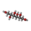
Mass: 182.172 Da / Num. of mol.: 1 / Source method: obtained synthetically / Formula: C6H14O6
#5: Chemical
ChemComp-IMD / IMIDAZOLE


Mass: 69.085 Da / Num. of mol.: 5 / Source method: obtained synthetically / Formula: C3H5N2
#6: Chemical ChemComp-ACT / ACETATE ION


Mass: 59.044 Da / Num. of mol.: 3 / Source method: obtained synthetically / Formula: C2H3O2
#7: Water ChemComp-HOH / water


Mass: 18.015 Da / Num. of mol.: 287 / Source method: isolated from a natural source / Formula: H2O

-
Experimental details

-
Experiment

ExperimentMethod: X-RAY DIFFRACTION / Number of used crystals: 1

-
Sample preparation

CrystalDensity Matthews: 1.98 Å3/Da / Density % sol: 37.78 %
Crystal growTemperature: 298 K / Method: vapor diffusion, sitting drop / pH: 8
Details: Protein (10 mM HEPES pH 7.5, 5 mM DTT, 10 mM L-sorbitol); Reservoir (MCSG4 A7)(0.2 M Zinc Acetate Dihydrate, 0.1 M Imidazole pH 8, 2.5 M Sodium Chloride); Cryoprotection (20% Diethylene Glycol, 80% Reservoir)

-
Data collection

DiffractionMean temperature: 100 K
Diffraction sourceSource: SYNCHROTRON / Site: APS / Beamline: 31-ID / Wavelength: 0.9793 Å
DetectorType: RAYONIX MX225HE / Detector: CCD / Date: Oct 20, 2015 / Details: MIRRORS
RadiationMonochromator: GRAPHITE / Protocol: SINGLE WAVELENGTH / Monochromatic (M) / Laue (L): M / Scattering type: x-ray
Radiation wavelengthWavelength: 0.9793 Å / Relative weight: 1
ReflectionResolution: 1.2→50 Å / Num. obs: 91508 / % possible obs: 99.9 % / Redundancy: 7.1 % / Rmerge(I) obs: 0.091 / Rpim(I) all: 0.037 / Rrim(I) all: 0.098 / Χ2: 1.262 / Net I/av σ(I): 24.165 / Net I/σ(I): 8.8 / Num. measured all: 650580
Reflection shell

Diffraction-ID: 1 / Rejects: _

Resolution (Å)Redundancy (%)Rmerge(I) obsNum. unique allCC1/2Rpim(I) allRrim(I) allΧ2% possible all
1.2-1.2470.68590210.8290.2770.741.03799.7
1.24-1.297.10.55790470.8790.2230.6011.049100
1.29-1.357.20.43790540.9220.1750.4711.089100
1.35-1.427.20.34690750.9460.1370.3721.139100
1.42-1.517.30.24790870.9720.0980.2661.237100
1.51-1.637.30.17690840.9850.070.191.255100
1.63-1.797.30.13791400.9890.0550.1481.441100
1.79-2.0570.11291810.9930.0460.1211.419100
2.05-2.5970.07692220.9960.0310.0821.442100
2.59-506.60.05495970.9970.0230.0581.5199.8

-
Processing

Software
NameVersionClassification
HKL-3000data scaling
REFMAC5.8.0123refinement
PDB_EXTRACT3.15data extraction
HKL-3000data reduction
MOLREPphasing
RefinementMethod to determine structure: MOLECULAR REPLACEMENT
Starting model: 4RS3
Resolution: 1.2→47.71 Å / Cor.coef. Fo:Fc: 0.972 / Cor.coef. Fo:Fc free: 0.966 / SU B: 0.963 / SU ML: 0.025 / Cross valid method: THROUGHOUT / σ(F): 0 / ESU R: 0.036 / ESU R Free: 0.038 / Stereochemistry target values: MAXIMUM LIKELIHOOD
Details: HYDROGENS HAVE BEEN ADDED IN THE RIDING POSITIONS U VALUES : WITH TLS ADDED
RfactorNum. reflection% reflectionSelection details
Rfree0.1779 4465 4.9 %RANDOM
Rwork0.1558 ---
obs0.1568 86963 99.95 %-
Solvent computationIon probe radii: 0.8 Å / Shrinkage radii: 0.8 Å / VDW probe radii: 1.2 Å / Solvent model: MASK
Displacement parametersBiso max: 57.35 Å2 / Biso mean: 14.242 Å2 / Biso min: 4.85 Å2
Baniso -1Baniso -2Baniso -3
1--0.15 Å2-0 Å2-0 Å2
2---0.21 Å20 Å2
3---0.36 Å2
Refinement stepCycle: final / Resolution: 1.2→47.71 Å
ProteinNucleic acidLigandSolventTotal
Num. atoms2308 0 73 287 2668
Biso mean--17.65 25.26 -
Num. residues----314
Refine LS restraints
Refine-IDTypeDev idealDev ideal targetNumber
X-RAY DIFFRACTIONr_bond_refined_d0.0360.022436
X-RAY DIFFRACTIONr_bond_other_d0.0020.022376
X-RAY DIFFRACTIONr_angle_refined_deg2.8281.9833302
X-RAY DIFFRACTIONr_angle_other_deg1.213.0015478
X-RAY DIFFRACTIONr_dihedral_angle_1_deg6.5345327
X-RAY DIFFRACTIONr_dihedral_angle_2_deg41.93827.327101
X-RAY DIFFRACTIONr_dihedral_angle_3_deg12.05115416
X-RAY DIFFRACTIONr_dihedral_angle_4_deg12.529157
X-RAY DIFFRACTIONr_chiral_restr0.1680.2385
X-RAY DIFFRACTIONr_gen_planes_refined0.0150.0212854
X-RAY DIFFRACTIONr_gen_planes_other0.0020.02501
X-RAY DIFFRACTIONr_mcbond_it1.2730.8731284
X-RAY DIFFRACTIONr_mcbond_other1.2420.8691280
X-RAY DIFFRACTIONr_mcangle_it1.8331.311608
LS refinement shellResolution: 1.2→1.231 Å / Total num. of bins used: 20
RfactorNum. reflection% reflection
Rfree0.245 340 -
Rwork0.213 6333 -
all-6673 -
obs--99.7 %
Refinement TLS params.Method: refined / Origin x: -3.9217 Å / Origin y: -23.338 Å / Origin z: 1.9355 Å
111213212223313233
T0.0085 Å20.0017 Å20.001 Å2-0.0185 Å2-0.001 Å2--0.0025 Å2
L0.582 °20.2019 °2-0.0234 °2-0.5282 °20.0687 °2--0.1709 °2
S-0.0177 Å °0.029 Å °-0.0068 Å °0.0058 Å °0.0235 Å °0.0155 Å °0.0152 Å °0.01 Å °-0.0058 Å °

+
About Yorodumi

-
News

-
Feb 9, 2022. New format data for meta-information of EMDB entries

New format data for meta-information of EMDB entries

  • Version 3 of the EMDB header file is now the official format.
  • The previous official version 1.9 will be removed from the archive.

Related info.:EMDB header

External links:wwPDB to switch to version 3 of the EMDB data model

-
Aug 12, 2020. Covid-19 info

Covid-19 info

URL: https://pdbjlvh1.pdbj.org/emnavi/covid19.php

New page: Covid-19 featured information page in EM Navigator.

Related info.:Covid-19 info / Mar 5, 2020. Novel coronavirus structure data

+
Mar 5, 2020. Novel coronavirus structure data

Novel coronavirus structure data

Related info.:Yorodumi Speices / Aug 12, 2020. Covid-19 info

External links:COVID-19 featured content - PDBj / Molecule of the Month (242):Coronavirus Proteases

+
Jan 31, 2019. EMDB accession codes are about to change! (news from PDBe EMDB page)

EMDB accession codes are about to change! (news from PDBe EMDB page)

  • The allocation of 4 digits for EMDB accession codes will soon come to an end. Whilst these codes will remain in use, new EMDB accession codes will include an additional digit and will expand incrementally as the available range of codes is exhausted. The current 4-digit format prefixed with “EMD-” (i.e. EMD-XXXX) will advance to a 5-digit format (i.e. EMD-XXXXX), and so on. It is currently estimated that the 4-digit codes will be depleted around Spring 2019, at which point the 5-digit format will come into force.
  • The EM Navigator/Yorodumi systems omit the EMD- prefix.

Related info.:Q: What is EMD? / ID/Accession-code notation in Yorodumi/EM Navigator

External links:EMDB Accession Codes are Changing Soon! / Contact to PDBj

+
Jul 12, 2017. Major update of PDB

Major update of PDB

  • wwPDB released updated PDB data conforming to the new PDBx/mmCIF dictionary.
  • This is a major update changing the version number from 4 to 5, and with Remediation, in which all the entries are updated.
  • In this update, many items about electron microscopy experimental information are reorganized (e.g. em_software).
  • Now, EM Navigator and Yorodumi are based on the updated data.

External links:wwPDB Remediation / Enriched Model Files Conforming to OneDep Data Standards Now Available in the PDB FTP Archive

-
Yorodumi

Thousand views of thousand structures

  • Yorodumi is a browser for structure data from EMDB, PDB, SASBDB, etc.
  • This page is also the successor to EM Navigator detail page, and also detail information page/front-end page for Omokage search.
  • The word "yorodu" (or yorozu) is an old Japanese word meaning "ten thousand". "mi" (miru) is to see.

Related info.:EMDB / PDB / SASBDB / Comparison of 3 databanks / Yorodumi Search / Aug 31, 2016. New EM Navigator & Yorodumi / Yorodumi Papers / Jmol/JSmol / Function and homology information / Changes in new EM Navigator and Yorodumi

Read more