| Entry | Database: PDB / ID: 5hde
|
|---|
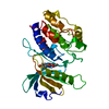
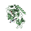
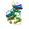
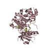
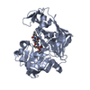
| Title | Crystal Structure of PTPN12 Catalytic Domain |
|---|
 Components Components | Tyrosine-protein phosphatase non-receptor type 12 |
|---|
 Keywords Keywords | HYDROLASE / PTPN12 / PTP / protein tyrosine phosphatase / dephosphorylation |
|---|
| Function / homology |  Function and homology information Function and homology information
negative regulation of platelet-derived growth factor receptor-beta signaling pathway / negative regulation of ERBB signaling pathway / regulation of epidermal growth factor receptor signaling pathway / tissue regeneration / Signaling by PDGF / Interleukin-37 signaling / podosome / protein dephosphorylation / peptidyl-tyrosine dephosphorylation / non-membrane spanning protein tyrosine phosphatase activity ...negative regulation of platelet-derived growth factor receptor-beta signaling pathway / negative regulation of ERBB signaling pathway / regulation of epidermal growth factor receptor signaling pathway / tissue regeneration / Signaling by PDGF / Interleukin-37 signaling / podosome / protein dephosphorylation / peptidyl-tyrosine dephosphorylation / non-membrane spanning protein tyrosine phosphatase activity / phosphoprotein phosphatase activity / protein-tyrosine-phosphatase / protein tyrosine phosphatase activity / SHC1 events in ERBB2 signaling / cellular response to epidermal growth factor stimulus / Constitutive Signaling by Overexpressed ERBB2 / EGFR downregulation / SH3 domain binding / Downregulation of ERBB2 signaling / focal adhesion / nucleoplasm / nucleus / cytoplasm / cytosolSimilarity search - Function Protein-tyrosine phosphatase, non-receptor type-12 / : / : / Protein tyrosine phosphatase superfamily / Protein-Tyrosine Phosphatase; Chain A / Protein tyrosine phosphatase, catalytic domain / PTP type protein phosphatase domain profile. / Protein-tyrosine phosphatase / Tyrosine-specific protein phosphatase, PTPase domain / Protein-tyrosine phosphatase, catalytic ...Protein-tyrosine phosphatase, non-receptor type-12 / : / : / Protein tyrosine phosphatase superfamily / Protein-Tyrosine Phosphatase; Chain A / Protein tyrosine phosphatase, catalytic domain / PTP type protein phosphatase domain profile. / Protein-tyrosine phosphatase / Tyrosine-specific protein phosphatase, PTPase domain / Protein-tyrosine phosphatase, catalytic / Protein tyrosine phosphatase, catalytic domain motif / Tyrosine specific protein phosphatases active site. / Protein-tyrosine phosphatase, active site / Tyrosine specific protein phosphatases domain profile. / Tyrosine-specific protein phosphatases domain / Protein-tyrosine phosphatase-like / Alpha-Beta Complex / Alpha BetaSimilarity search - Domain/homology |
|---|
| Biological species |  Homo sapiens (human) Homo sapiens (human) |
|---|
| Method |  X-RAY DIFFRACTION / SYNCHROTRON / MOLECULAR REPLACEMENT / Resolution: 1.62 Å X-RAY DIFFRACTION / SYNCHROTRON / MOLECULAR REPLACEMENT / Resolution: 1.62 Å |
|---|
 Authors Authors | Dong, H. / Li, S. / Shi, J. |
|---|
| Funding support |  China, 2items China, 2items | Organization | Grant number | Country |
|---|
| the National Natural Science Foundation of China | 31300601 | China | | the PUMC Youth Fund | 33320140186 | China |
|
|---|
 Citation Citation | Journal: To Be Published
Title: Crystal Structure of PTPN12 Catalytic Domain
Authors: Dong, H. / Shi, J. / Li, J. / Li, S. |
|---|
| History | | Deposition | Jan 5, 2016 | Deposition site: RCSB / Processing site: PDBJ |
|---|
| Revision 1.0 | Jan 18, 2017 | Provider: repository / Type: Initial release |
|---|
| Revision 1.1 | Oct 4, 2017 | Group: Data collection / Category: diffrn_detector / Item: _diffrn_detector.detector |
|---|
| Revision 1.2 | Oct 11, 2017 | Group: Data collection / Category: diffrn_source
Item: _diffrn_source.pdbx_synchrotron_beamline / _diffrn_source.pdbx_synchrotron_site / _diffrn_source.source |
|---|
| Revision 1.3 | Nov 8, 2023 | Group: Data collection / Database references / Refinement description
Category: chem_comp_atom / chem_comp_bond ...chem_comp_atom / chem_comp_bond / database_2 / pdbx_initial_refinement_model
Item: _database_2.pdbx_DOI / _database_2.pdbx_database_accession |
|---|
| Revision 1.4 | Sep 17, 2025 | Group: Derived calculations / Structure summary
Category: pdbx_entry_details / pdbx_modification_feature / struct_conn |
|---|
|
|---|
 Movie
Movie Controller


 Open data
 Basic information
 Structure visualization
 Downloads & links
 Other downloads
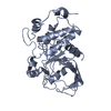


 PDBj
PDBj


 Assembly
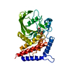


 Mass: 94.971 Da / Num. of mol.: 1 / Source method: obtained synthetically / Formula: PO4
 Mass: 18.015 Da / Num. of mol.: 354 / Source method: isolated from a natural source / Formula: H2O
 Sample preparation
 / Beamline: BL-5A / Wavelength: 1 Å
 Processing