- PDB-5ha9: Crystal structure-based design and disovery of a novel PARP1 anti... -
+
Open data
ID or keywords:
Loading...
-
Basic information
Entry
Database: PDB / ID: 5ha9
Title
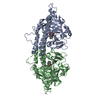
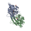
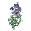
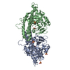
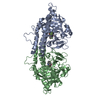
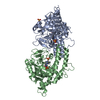
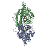
Crystal structure-based design and disovery of a novel PARP1 antiagonist (BL-PA10) that induces apoptosis and inhibits metastasis in triple negative breast cancer
Protocol: SINGLE WAVELENGTH / Monochromatic (M) / Laue (L): M / Scattering type: x-ray
Radiation wavelength
Wavelength: 1.006323 Å / Relative weight: 1
Reflection
Resolution: 4→50 Å / Num. obs: 6434 / % possible obs: 99.1 % / Redundancy: 4.5 % / Rmerge(I) obs: 0.212 / Χ2: 2.04 / Net I/av σ(I): 9.947 / Net I/σ(I): 6.2 / Num. measured all: 29162
Reflection shell
Diffraction-ID: 1 / Rejects: _
Resolution (Å)
Redundancy (%)
Rmerge(I) obs
Num. unique all
Χ2
% possible all
4-4.07
4.8
0.398
292
1.962
100
4.07-4.14
4.7
0.437
322
1.94
99.7
4.14-4.22
4.7
0.349
304
2.106
100
4.22-4.31
4.5
0.348
325
2.207
98.5
4.31-4.4
4.5
0.291
307
2.577
98.4
4.4-4.5
4.8
0.286
313
2.179
99.4
4.5-4.62
4.6
0.253
320
2.324
100
4.62-4.74
4.6
0.258
319
2.249
99.7
4.74-4.88
4.6
0.24
315
2.411
99.4
4.88-5.04
4.7
0.238
306
2.123
99.7
5.04-5.22
4.6
0.267
323
1.947
99.7
5.22-5.43
4.4
0.256
324
1.93
99.1
5.43-5.67
4.7
0.297
318
1.897
100
5.67-5.97
4.4
0.272
331
1.689
99.7
5.97-6.35
4.5
0.255
310
1.606
98.4
6.35-6.84
4.1
0.218
336
1.573
99.7
6.84-7.52
4.7
0.14
320
1.591
99.7
7.52-8.6
4.5
0.109
341
1.784
99.7
8.6-10.82
4.4
0.082
342
2.12
99.4
10.82-50
3.8
0.086
366
2.651
93.8
-
Processing
Software
Name
Version
Classification
HKL-2000
datascaling
REFMAC
5.8.0049
refinement
PDB_EXTRACT
3.15
dataextraction
MOLREP
phasing
Refinement
Resolution: 4.01→50 Å / Cor.coef. Fo:Fc: 0.888 / Cor.coef. Fo:Fc free: 0.73 / SU B: 101.143 / SU ML: 1.276 / Cross valid method: THROUGHOUT / σ(F): 0 / ESU R Free: 1.399 / Stereochemistry target values: MAXIMUM LIKELIHOOD Details: HYDROGENS HAVE BEEN ADDED IN THE RIDING POSITIONS U VALUES : REFINED INDIVIDUALLY
Rfactor
Num. reflection
% reflection
Selection details
Rfree
0.3578
300
4.7 %
RANDOM
Rwork
0.2421
-
-
-
obs
0.2475
6104
98.31 %
-
Solvent computation
Ion probe radii: 0.8 Å / Shrinkage radii: 0.8 Å / VDW probe radii: 1.2 Å / Solvent model: MASK
In the structure databanks used in Yorodumi, some data are registered as the other names, "COVID-19 virus" and "2019-nCoV". Here are the details of the virus and the list of structure data.
Jan 31, 2019. EMDB accession codes are about to change! (news from PDBe EMDB page)
EMDB accession codes are about to change! (news from PDBe EMDB page)
The allocation of 4 digits for EMDB accession codes will soon come to an end. Whilst these codes will remain in use, new EMDB accession codes will include an additional digit and will expand incrementally as the available range of codes is exhausted. The current 4-digit format prefixed with “EMD-” (i.e. EMD-XXXX) will advance to a 5-digit format (i.e. EMD-XXXXX), and so on. It is currently estimated that the 4-digit codes will be depleted around Spring 2019, at which point the 5-digit format will come into force.
The EM Navigator/Yorodumi systems omit the EMD- prefix.
Related info.:Q: What is EMD? / ID/Accession-code notation in Yorodumi/EM Navigator
Yorodumi is a browser for structure data from EMDB, PDB, SASBDB, etc.
This page is also the successor to EM Navigator detail page, and also detail information page/front-end page for Omokage search.
The word "yorodu" (or yorozu) is an old Japanese word meaning "ten thousand". "mi" (miru) is to see.
Related info.:EMDB / PDB / SASBDB / Comparison of 3 databanks / Yorodumi Search / Aug 31, 2016. New EM Navigator & Yorodumi / Yorodumi Papers / Jmol/JSmol / Function and homology information / Changes in new EM Navigator and Yorodumi
 Movie
Movie Controller

 Yorodumi
 Open data
 Basic information
 Components
 Keywords
 Function and homology information
 Homo sapiens (human)
 X-RAY DIFFRACTION /
  Authors
 Citation
 Structure visualization
 Downloads & links
 Other downloads
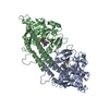


 PDBj
PDBj


 Assembly
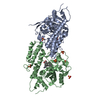


 Mass: 92.094 Da / Num. of mol.: 2 / Source method: obtained synthetically / Formula: C3H8O3
 Mass: 96.063 Da / Num. of mol.: 6 / Source method: obtained synthetically / Formula: SO4
 Mass: 277.403 Da / Num. of mol.: 1 / Source method: obtained synthetically / Formula: C20H23N
 Sample preparation
 / Beamline: BL17U / Wavelength: 1.006323 Å
 Processing