 Movie
Movie Controller

[English] 日本語
 Yorodumi
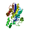
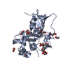
- PDB-5h6n: DNA targeting ADP-ribosyltransferase Pierisin-1, autoinhibitory form -
+ Open data

- Basic information
| Entry | Database: PDB / ID: 5h6n | ||||||
|---|---|---|---|---|---|---|---|
| Title | DNA targeting ADP-ribosyltransferase Pierisin-1, autoinhibitory form | ||||||
 Components Components | Pierisin-1 | ||||||
 Keywords Keywords | TRANSFERASE / DNA-targeting ADP-ribosyltransferase | ||||||
| Function / homology |  Function and homology information Function and homology information2'-deoxyguanosine DNA ADP-ribosyltransferase activity / DNA ADP-ribosylation / glycosyltransferase activity / Transferases; Glycosyltransferases; Pentosyltransferases / nucleotidyltransferase activity / apoptotic process / DNA binding Similarity search - Function | ||||||
| Biological species |  Pieris rapae (cabbage white) Pieris rapae (cabbage white) | ||||||
| Method |  X-RAY DIFFRACTION / SYNCHROTRON / MOLECULAR REPLACEMENT / Resolution: 1.8 Å X-RAY DIFFRACTION / SYNCHROTRON / MOLECULAR REPLACEMENT / Resolution: 1.8 Å | ||||||
 Authors Authors | Oda, T. / Hirabayashi, H. / Shikauchi, G. / Takamura, R. / Hiraga, K. / Minami, H. / Hashimoto, H. / Yamamoto, M. / Wakabayashi, K. / Sugimura, T. ...Oda, T. / Hirabayashi, H. / Shikauchi, G. / Takamura, R. / Hiraga, K. / Minami, H. / Hashimoto, H. / Yamamoto, M. / Wakabayashi, K. / Sugimura, T. / Shimizu, T. / Sato, M. | ||||||
 Citation Citation | Journal: J. Biol. Chem. / Year: 2017 Title: Structural basis of autoinhibition and activation of the DNA-targeting ADP-ribosyltransferase pierisin-1 Authors: Oda, T. / Hirabayashi, H. / Shikauchi, G. / Takamura, R. / Hiraga, K. / Minami, H. / Hashimoto, H. / Yamamoto, M. / Wakabayashi, K. / Shimizu, T. / Sato, M. | ||||||
| History |
|
- Structure visualization
| Structure viewer | Molecule: MolmilJmol/JSmol |
|---|
- Downloads & links
-Download
| PDBx/mmCIF format | 5h6n.cif.gz | 214.8 KB | Display | PDBx/mmCIF format |
|---|---|---|---|---|
| PDB format | pdb5h6n.ent.gz | 171.6 KB | Display | PDB format |
| PDBx/mmJSON format | 5h6n.json.gz | Tree view | PDBx/mmJSON format | |
| Others |  Other downloads Other downloads |
-Validation report
| Arichive directory | https://data.pdbj.org/pub/pdb/validation_reports/h6/5h6nftp://data.pdbj.org/pub/pdb/validation_reports/h6/5h6n | HTTPS FTP |
|---|
-Related structure data
| Related structure data |  5h6jC  5h6kC 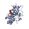 5h6lC  5h6mC  2cb4S C: citing same article ( S: Starting model for refinement |
|---|---|
| Similar structure data |


-Links
 PDBj
PDBj

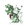
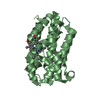
- Assembly
| Deposited unit | 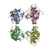
| ||||||||
|---|---|---|---|---|---|---|---|---|---|
| 1 | 
| ||||||||
| 2 | 
| ||||||||
| 3 | 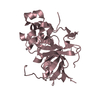
| ||||||||
| 4 | 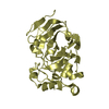
| ||||||||
| Unit cell |
|
-Components
| #1: Protein | Mass: 31456.125 Da / Num. of mol.: 4 / Fragment: UNP residues 1-267 / Mutation: E165Q Source method: isolated from a genetically manipulated source Source: (gene. exp.) Pieris rapae (cabbage white) / Plasmid: pGEX6p-1 / Production host:  References: UniProt: H3JU00, UniProt: Q9U8Q4*PLUS, Transferases; Glycosyltransferases; Pentosyltransferases #2: Water | ChemComp-HOH / |  Mass: 18.015 Da / Num. of mol.: 588 / Source method: isolated from a natural source / Formula: H2O Mass: 18.015 Da / Num. of mol.: 588 / Source method: isolated from a natural source / Formula: H2O |
|---|
-Experimental details
-Experiment
| Experiment | Method: X-RAY DIFFRACTION / Number of used crystals: 1 |
|---|
- Sample preparation
| Crystal | Density Matthews: 3.06 Å3/Da / Density % sol: 59.78 % |
|---|---|
| Crystal grow | Temperature: 293 K / Method: vapor diffusion, hanging drop / pH: 8 / Details: 22% (v/v) t-butanol and 0.1M Tris-HCl pH8 |
-Data collection
| Diffraction | Mean temperature: 93 K |
|---|---|
| Diffraction source | Source: SYNCHROTRON / Site: Photon Factory  / Beamline: BL-5A / Wavelength: 1 Å / Beamline: BL-5A / Wavelength: 1 Å |
| Detector | Type: ADSC QUANTUM 315 / Detector: CCD / Date: Oct 7, 2007 |
| Radiation | Protocol: SINGLE WAVELENGTH / Monochromatic (M) / Laue (L): M / Scattering type: x-ray |
| Radiation wavelength | Wavelength: 1 Å / Relative weight: 1 |
| Reflection | Resolution: 1.8→50 Å / Num. obs: 132451 / % possible obs: 95 % / Redundancy: 3.74 % / Net I/σ(I): 19.3 |
- Processing
| Software |
| ||||||||||||||||||||||||||||||||||||||||||||||||||||||||||||||||||||||||||||||||||||||||||||||||||||||||||||||||||||||||||||||||||||||||||||||||||||||||||||||||||||||||||||||||||||||
|---|---|---|---|---|---|---|---|---|---|---|---|---|---|---|---|---|---|---|---|---|---|---|---|---|---|---|---|---|---|---|---|---|---|---|---|---|---|---|---|---|---|---|---|---|---|---|---|---|---|---|---|---|---|---|---|---|---|---|---|---|---|---|---|---|---|---|---|---|---|---|---|---|---|---|---|---|---|---|---|---|---|---|---|---|---|---|---|---|---|---|---|---|---|---|---|---|---|---|---|---|---|---|---|---|---|---|---|---|---|---|---|---|---|---|---|---|---|---|---|---|---|---|---|---|---|---|---|---|---|---|---|---|---|---|---|---|---|---|---|---|---|---|---|---|---|---|---|---|---|---|---|---|---|---|---|---|---|---|---|---|---|---|---|---|---|---|---|---|---|---|---|---|---|---|---|---|---|---|---|---|---|---|---|
| Refinement | Method to determine structure: MOLECULAR REPLACEMENT Starting model: 2CB4 Resolution: 1.8→30.068 Å / Cor.coef. Fo:Fc: 0.937 / Cor.coef. Fo:Fc free: 0.927 / SU B: 2.909 / SU ML: 0.088 / Cross valid method: THROUGHOUT / ESU R: 0.127 / ESU R Free: 0.117 / Stereochemistry target values: MAXIMUM LIKELIHOOD / Details: HYDROGENS HAVE BEEN ADDED IN THE RIDING POSITIONS
| ||||||||||||||||||||||||||||||||||||||||||||||||||||||||||||||||||||||||||||||||||||||||||||||||||||||||||||||||||||||||||||||||||||||||||||||||||||||||||||||||||||||||||||||||||||||
| Solvent computation | Ion probe radii: 0.8 Å / Shrinkage radii: 0.8 Å / VDW probe radii: 1.2 Å / Solvent model: MASK | ||||||||||||||||||||||||||||||||||||||||||||||||||||||||||||||||||||||||||||||||||||||||||||||||||||||||||||||||||||||||||||||||||||||||||||||||||||||||||||||||||||||||||||||||||||||
| Displacement parameters | Biso mean: 31.354 Å2
| ||||||||||||||||||||||||||||||||||||||||||||||||||||||||||||||||||||||||||||||||||||||||||||||||||||||||||||||||||||||||||||||||||||||||||||||||||||||||||||||||||||||||||||||||||||||
| Refinement step | Cycle: 1 / Resolution: 1.8→30.068 Å
| ||||||||||||||||||||||||||||||||||||||||||||||||||||||||||||||||||||||||||||||||||||||||||||||||||||||||||||||||||||||||||||||||||||||||||||||||||||||||||||||||||||||||||||||||||||||
| Refine LS restraints |
|