 Movie
Movie Controller

[English] 日本語
 Yorodumi
- PDB-5h5e: The crystal structure of the yeast arginine methyltransferase SFM... -
+ Open data

- Basic information
| Entry | Database: PDB / ID: 5h5e | ||||||
|---|---|---|---|---|---|---|---|
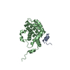
| Title | The crystal structure of the yeast arginine methyltransferase SFM1 complexed with SAH | ||||||
 Components Components | Protein arginine N-methyltransferase SFM1 | ||||||
 Keywords Keywords | TRANSFERASE / arginine methyltransferase / spout fold | ||||||
| Function / homology | Protein arginine N-methyltransferase SFM1-like / Protein arginine N-methyltransferase SFM1-like / protein-arginine omega-N monomethyltransferase activity / Transferases; Transferring one-carbon groups; Methyltransferases / methylation / cytoplasm / S-ADENOSYL-L-HOMOCYSTEINE / Protein arginine N-methyltransferase SFM1 Function and homology information Function and homology information | ||||||
| Biological species |  | ||||||
| Method |  X-RAY DIFFRACTION / SYNCHROTRON / MOLECULAR REPLACEMENT / Resolution: 2.09 Å X-RAY DIFFRACTION / SYNCHROTRON / MOLECULAR REPLACEMENT / Resolution: 2.09 Å | ||||||
 Authors Authors | Xie, W. / Wang, C. / Zeng, J. | ||||||
 Citation Citation | Journal: FEBS Lett. / Year: 2017 Title: A flexible cofactor-binding loop in the novel arginine methyltransferase Sfm1. Authors: Wang, C. / Zeng, J. / Xie, W. | ||||||
| History |
|
- Structure visualization
| Structure viewer | Molecule: MolmilJmol/JSmol |
|---|
- Downloads & links
-Download
| PDBx/mmCIF format | 5h5e.cif.gz | 59.5 KB | Display | PDBx/mmCIF format |
|---|---|---|---|---|
| PDB format | pdb5h5e.ent.gz | 40.8 KB | Display | PDB format |
| PDBx/mmJSON format | 5h5e.json.gz | Tree view | PDBx/mmJSON format | |
| Others |  Other downloads Other downloads |
-Validation report
| Arichive directory | https://data.pdbj.org/pub/pdb/validation_reports/h5/5h5eftp://data.pdbj.org/pub/pdb/validation_reports/h5/5h5e | HTTPS FTP |
|---|
-Related structure data
| Related structure data | 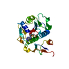 5h5dC 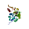 5h5fC 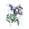 5c77S C: citing same article ( S: Starting model for refinement |
|---|---|
| Similar structure data |


-Links
 PDBj
PDBj- Assembly
| Deposited unit | 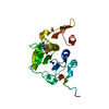
| ||||||||
|---|---|---|---|---|---|---|---|---|---|
| 1 |
| ||||||||
| Unit cell |
|
-Components
| #1: Protein | Mass: 27148.129 Da / Num. of mol.: 1 Source method: isolated from a genetically manipulated source Source: (gene. exp.) Strain: ATCC 204508 / S288c / Gene: SFM1, YOR021C, OR26.11 / Production host:  References: UniProt: Q12314, Transferases; Transferring one-carbon groups; Methyltransferases |
|---|---|
| #2: Chemical | ChemComp-SAH /   Type: L-peptide linking / Mass: 384.411 Da / Num. of mol.: 1 / Source method: obtained synthetically / Formula: C14H20N6O5S Type: L-peptide linking / Mass: 384.411 Da / Num. of mol.: 1 / Source method: obtained synthetically / Formula: C14H20N6O5S |
| #3: Water | ChemComp-HOH /  Mass: 18.015 Da / Num. of mol.: 47 / Source method: isolated from a natural source / Formula: H2O Mass: 18.015 Da / Num. of mol.: 47 / Source method: isolated from a natural source / Formula: H2O |
-Experimental details
-Experiment
| Experiment | Method: X-RAY DIFFRACTION / Number of used crystals: 1 |
|---|
- Sample preparation
| Crystal | Density Matthews: 1.72 Å3/Da / Density % sol: 28.57 % |
|---|---|
| Crystal grow | Temperature: 298 K / Method: vapor diffusion, sitting drop / pH: 8.5 / Details: 0.2 M NaCl, 0.1 M Tris-HCl (pH 8.5), 38% PEG 3350 |
-Data collection
| Diffraction | Mean temperature: 100 K |
|---|---|
| Diffraction source | Source: SYNCHROTRON / Site: SSRF  / Beamline: BL17U / Wavelength: 0.979 Å / Beamline: BL17U / Wavelength: 0.979 Å |
| Detector | Type: ADSC QUANTUM 4 / Detector: CCD / Date: Jul 13, 2016 |
| Radiation | Protocol: SINGLE WAVELENGTH / Monochromatic (M) / Laue (L): M / Scattering type: x-ray |
| Radiation wavelength | Wavelength: 0.979 Å / Relative weight: 1 |
| Reflection | Resolution: 2.1→50 Å / Num. obs: 10918 / % possible obs: 99.7 % / Redundancy: 3.7 % / Rmerge(I) obs: 0.089 / Net I/σ(I): 23.7 |
| Reflection shell | Resolution: 2.1→2.18 Å / Redundancy: 3.7 % / Rmerge(I) obs: 0.496 / Mean I/σ(I) obs: 3.4 / CC1/2: 0.84 / % possible all: 99.5 |
- Processing
| Software |
| |||||||||||||||||||||||||||||||||||
|---|---|---|---|---|---|---|---|---|---|---|---|---|---|---|---|---|---|---|---|---|---|---|---|---|---|---|---|---|---|---|---|---|---|---|---|---|
| Refinement | Method to determine structure: MOLECULAR REPLACEMENT Starting model: 5C77 Resolution: 2.09→37.155 Å / SU ML: 0.23 / Cross valid method: FREE R-VALUE / σ(F): 1.35 / Phase error: 25.4 / Stereochemistry target values: ML
| |||||||||||||||||||||||||||||||||||
| Solvent computation | Shrinkage radii: 0.9 Å / VDW probe radii: 1.11 Å / Solvent model: FLAT BULK SOLVENT MODEL | |||||||||||||||||||||||||||||||||||
| Refinement step | Cycle: LAST / Resolution: 2.09→37.155 Å
| |||||||||||||||||||||||||||||||||||
| Refine LS restraints |
| |||||||||||||||||||||||||||||||||||
| LS refinement shell |
|
