 Movie
Movie Controller

[English] 日本語
 Yorodumi
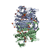
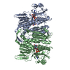
- PDB-5grt: HUMAN GLUTATHIONE REDUCTASE A34E, R37W MUTANT, GLUTATHIONYLSPERMI... -
+ Open data

- Basic information
| Entry | Database: PDB / ID: 5grt | ||||||
|---|---|---|---|---|---|---|---|
| Title | HUMAN GLUTATHIONE REDUCTASE A34E, R37W MUTANT, GLUTATHIONYLSPERMIDINE COMPLEX | ||||||
 Components Components | GLUTATHIONE REDUCTASE | ||||||
 Keywords Keywords | OXIDOREDUCTASE / FLAVOENZYME / GLUTATHIONYL SPERMIDINE | ||||||
| Function / homology |  Function and homology information Function and homology informationglutathione-disulfide reductase / Metabolism of ingested H2SeO4 and H2SeO3 into H2Se / glutathione-disulfide reductase (NADPH) activity / Interconversion of nucleotide di- and triphosphates / NFE2L2 regulating anti-oxidant/detoxification enzymes / Detoxification of Reactive Oxygen Species / cell redox homeostasis / TP53 Regulates Metabolic Genes / glutathione metabolic process / NADP binding ...glutathione-disulfide reductase / Metabolism of ingested H2SeO4 and H2SeO3 into H2Se / glutathione-disulfide reductase (NADPH) activity / Interconversion of nucleotide di- and triphosphates / NFE2L2 regulating anti-oxidant/detoxification enzymes / Detoxification of Reactive Oxygen Species / cell redox homeostasis / TP53 Regulates Metabolic Genes / glutathione metabolic process / NADP binding / flavin adenine dinucleotide binding / cellular response to oxidative stress / electron transfer activity / mitochondrial matrix / external side of plasma membrane / mitochondrion / extracellular exosome / cytosol Similarity search - Function | ||||||
| Biological species |  Homo sapiens (human) Homo sapiens (human) | ||||||
| Method |  X-RAY DIFFRACTION / DIRECT BASED ON KNOWN MODEL / Resolution: 2.4 Å X-RAY DIFFRACTION / DIRECT BASED ON KNOWN MODEL / Resolution: 2.4 Å | ||||||
 Authors Authors | Stoll, V.S. / Simpson, S.J. / Krauth-Siegel, R.L. / Walsh, C.T. / Pai, E.F. | ||||||
 Citation Citation | Journal: Biochemistry / Year: 1997 Title: Glutathione reductase turned into trypanothione reductase: structural analysis of an engineered change in substrate specificity. Authors: Stoll, V.S. / Simpson, S.J. / Krauth-Siegel, R.L. / Walsh, C.T. / Pai, E.F. #1: Journal: Biochemistry / Year: 1991Title: Redox Enzyme Engineering: Conversion of Human Glutathione Reductase Into a Trypanothione Reductase Authors: Bradley, M. / Bucheler, U.S. / Walsh, C.T. #2: Journal: J.Mol.Biol. / Year: 1987Title: Refined Structure of Glutathione Reductase at 1.54 A Resolution Authors: Karplus, P.A. / Schulz, G.E. #3: Journal: J.Biol.Chem. / Year: 1983Title: The Catalytic Mechanism of Glutathione Reductase as Derived from X-Ray Diffraction Analyses of Reaction Intermediates Authors: Pai, E.F. / Schulz, G.E. #4: Journal: J.Mol.Biol. / Year: 1983Title: Comparison of the Three-Dimensional Protein and Nucleotide Structure of the Fad-Binding Domain of P-Hydroxybenzoate Hydroxylase with the Fad-as Well as Nadph-Binding Domains of Glutathione Reductase Authors: Wierenga, R.K. / Drenth, J. / Schulz, G.E. #5: Journal: Eur.J.Biochem. / Year: 1982Title: Glutathione Reductase from Human Erythrocytes. The Sequences of the Nadph Domain and of the Interface Domain Authors: Krauth-Siegel, R.L. / Blatterspiel, R. / Saleh, M. / Schiltz, E. / Schirmer, R.H. / Untucht-Grau, R. #6: Journal: J.Mol.Biol. / Year: 1982Title: Fad-Binding Site of Glutathione Reductase Authors: Schulz, G.E. / Schirmer, R.H. / Pai, E.F. #7: Journal: J.Mol.Biol. / Year: 1981Title: Three-Dimensional Structure of Glutathione Reductase at 2 A Resolution Authors: Thieme, R. / Pai, E.F. / Schirmer, R.H. / Schulz, G.E. #8: Journal: J.Mol.Biol. / Year: 1980Title: Gene Duplication in Glutathione Reductase Authors: Schulz, G.E. #9: Journal: FEBS Lett. / Year: 1979Title: The C-Terminal Fragment of Human Glutathione Reductase Contains the Postulated Catalytic Histidine Authors: Untucht-Grau, R. / Schulz, G.E. / Schirmer, R.H. #10: Journal: Nature / Year: 1978Title: The Structure of the Flavoenzyme Glutathione Reductase Authors: Schulz, G.E. / Schirmer, R.H. / Sachsenheimer, W. / Pai, E.F. #11: Journal: J.Mol.Biol. / Year: 1977Title: Low Resolution Structure of Human Erythrocyte Glutathione Reductase Authors: Zappe, H.A. / Krohne-Ehrich, G. / Schulz, G.E. #12: Journal: FEBS Lett. / Year: 1975Title: Crystals of Human Erythrocyte Glutathione Reductase Authors: Schulz, G.E. / Zappe, H. / Worthington, D.J. / Rosemeyer, M.A. | ||||||
| History |
|
- Structure visualization
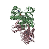
| Structure viewer | Molecule: MolmilJmol/JSmol |
|---|
- Downloads & links
-Download
| PDBx/mmCIF format | 5grt.cif.gz | 105.3 KB | Display | PDBx/mmCIF format |
|---|---|---|---|---|
| PDB format | pdb5grt.ent.gz | 78.5 KB | Display | PDB format |
| PDBx/mmJSON format | 5grt.json.gz | Tree view | PDBx/mmJSON format | |
| Others |  Other downloads Other downloads |
-Validation report
| Arichive directory | https://data.pdbj.org/pub/pdb/validation_reports/gr/5grtftp://data.pdbj.org/pub/pdb/validation_reports/gr/5grt | HTTPS FTP |
|---|
-Related structure data
| Related structure data |  1grtSC  2grtC  3grtC  4grtC S: Starting model for refinement C: citing same article ( |
|---|---|
| Similar structure data |


-Links
 PDBj
PDBj


- Assembly
| Deposited unit | 
| ||||||||
|---|---|---|---|---|---|---|---|---|---|
| 1 | 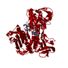
| ||||||||
| Unit cell |
|
-Components
| #1: Protein | Mass: 50065.480 Da / Num. of mol.: 1 / Mutation: A34E, R37W Source method: isolated from a genetically manipulated source Details: CONTAINS A NON-COVALENTLY BOUND FAD AND OXIDIZED GLUTATHIONYLSPERMIDINE SUBSTRATE Source: (gene. exp.) Homo sapiens (human) / Cell: RED BLOOD CELLS / Organ: BLOOD / Plasmid: PUB302 / Production host:  |
|---|---|
| #2: Chemical | ChemComp-FAD /   Mass: 785.550 Da / Num. of mol.: 1 / Source method: obtained synthetically / Formula: C27H33N9O15P2 / Comment: FAD*YM Mass: 785.550 Da / Num. of mol.: 1 / Source method: obtained synthetically / Formula: C27H33N9O15P2 / Comment: FAD*YM |
| #3: Chemical | ChemComp-TS4 /   Mass: 867.092 Da / Num. of mol.: 1 / Source method: obtained synthetically / Formula: C34H66N12O10S2 Mass: 867.092 Da / Num. of mol.: 1 / Source method: obtained synthetically / Formula: C34H66N12O10S2 |
| Has protein modification | Y |
-Experimental details
-Experiment
| Experiment | Method: X-RAY DIFFRACTION / Number of used crystals: 1 |
|---|
- Sample preparation
| Crystal | Density Matthews: 2.76 Å3/Da / Density % sol: 55.38 % | ||||||||||||||||||||||||||||||
|---|---|---|---|---|---|---|---|---|---|---|---|---|---|---|---|---|---|---|---|---|---|---|---|---|---|---|---|---|---|---|---|
| Crystal grow | pH: 8 Details: 0.57-0.90 M AMMONIUM SULFATE, 100 MM POTASSIUM PHOSPHATE, PH 8.0, AND 0.5% 1-N-BETA-OCTYL-D-GLUCOPYRANOSIDE HANGING DROP VAPOR DIFFUSION, CRYSTAL SOAKED IN ARTIFICIAL MOTHER LIQUOR AT PH 8. ...Details: 0.57-0.90 M AMMONIUM SULFATE, 100 MM POTASSIUM PHOSPHATE, PH 8.0, AND 0.5% 1-N-BETA-OCTYL-D-GLUCOPYRANOSIDE HANGING DROP VAPOR DIFFUSION, CRYSTAL SOAKED IN ARTIFICIAL MOTHER LIQUOR AT PH 8.0,CONTAINING 0.5% BETA-OCTYL GLUCOSIDE AND 42 MM GLUTATHIONYLSPERMIDINE. | ||||||||||||||||||||||||||||||
| Crystal grow | *PLUS Method: vapor diffusion, hanging drop | ||||||||||||||||||||||||||||||
| Components of the solutions | *PLUS
|
-Data collection
| Diffraction | Mean temperature: 277 K |
|---|---|
| Diffraction source | Source: ROTATING ANODE / Type: ELLIOTT GX-18 / Wavelength: 1.5418 |
| Detector | Type: SIEMENS / Detector: AREA DETECTOR / Date: Feb 1, 1992 / Details: MIRRORS |
| Radiation | Monochromator: NI FILTER / Monochromatic (M) / Laue (L): M / Scattering type: x-ray |
| Radiation wavelength | Wavelength: 1.5418 Å / Relative weight: 1 |
| Reflection | Highest resolution: 2.3 Å / Num. obs: 16124 / % possible obs: 84.8 % / Observed criterion σ(I): 0.1 / Redundancy: 1.8 % / Rsym value: 0.056 |
| Reflection shell | Resolution: 2.4→2.53 Å / % possible all: 40.9 |
| Reflection | *PLUS Num. measured all: 29646 / Rmerge(I) obs: 0.056 |
| Reflection shell | *PLUS % possible obs: 40.9 % |
- Processing
| Software |
| ||||||||||||||||||||||||||||||||||||||||||||||||||||||||||||
|---|---|---|---|---|---|---|---|---|---|---|---|---|---|---|---|---|---|---|---|---|---|---|---|---|---|---|---|---|---|---|---|---|---|---|---|---|---|---|---|---|---|---|---|---|---|---|---|---|---|---|---|---|---|---|---|---|---|---|---|---|---|
| Refinement | Method to determine structure: DIRECT BASED ON KNOWN MODEL Starting model: PDB ENTRY 1GRT Resolution: 2.4→10 Å / Data cutoff high absF: 10000000 / σ(F): 0.1
| ||||||||||||||||||||||||||||||||||||||||||||||||||||||||||||
| Refine analyze | Luzzati d res low obs: 10 Å | ||||||||||||||||||||||||||||||||||||||||||||||||||||||||||||
| Refinement step | Cycle: LAST / Resolution: 2.4→10 Å
| ||||||||||||||||||||||||||||||||||||||||||||||||||||||||||||
| Refine LS restraints |
| ||||||||||||||||||||||||||||||||||||||||||||||||||||||||||||
| LS refinement shell | Resolution: 2.4→2.53 Å / Total num. of bins used: 8
| ||||||||||||||||||||||||||||||||||||||||||||||||||||||||||||
| Xplor file |
| ||||||||||||||||||||||||||||||||||||||||||||||||||||||||||||
| Software | *PLUS Name: X-PLOR / Version: 3.1F / Classification: refinement | ||||||||||||||||||||||||||||||||||||||||||||||||||||||||||||
| Refinement | *PLUS Num. reflection obs: 16124 | ||||||||||||||||||||||||||||||||||||||||||||||||||||||||||||
| Solvent computation | *PLUS | ||||||||||||||||||||||||||||||||||||||||||||||||||||||||||||
| Displacement parameters | *PLUS | ||||||||||||||||||||||||||||||||||||||||||||||||||||||||||||
| Refine LS restraints | *PLUS
| ||||||||||||||||||||||||||||||||||||||||||||||||||||||||||||
| LS refinement shell | *PLUS Rfactor obs: 0.197 |
