 Movie
Movie Controller

[English] 日本語
 Yorodumi
- PDB-5fr0: The details of glycolipid glycan hydrolysis by the structural ana... -
+ Open data

- Basic information
| Entry | Database: PDB / ID: 5fr0 | ||||||
|---|---|---|---|---|---|---|---|
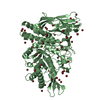
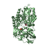
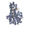
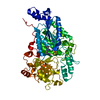
| Title | The details of glycolipid glycan hydrolysis by the structural analysis of a family 123 glycoside hydrolase from Clostridium perfringens | ||||||
 Components Components | BETA-N-ACETYLGALACTOSAMINIDASE | ||||||
 Keywords Keywords | HYDROLASE / GLYCOSIDE HYDROLASE / GH123 / GLYCOSPHINGOLIPID / GANGLIOSIDE / GLOBOSIDE / SUBSTRATE-ASSISTED CATALYSIS. | ||||||
| Function / homology | Glycoside hydrolase 123, N-terminal domain / : / Glycoside hydrolase 123 N-terminal domain / Glycoside hydrolase 123, C-terminal / Glycoside hydrolase 123, catalytic domain / PHOSPHATE ION / Chem-SIZ / Uncharacterized protein Function and homology information Function and homology information | ||||||
| Biological species |   CLOSTRIDIUM PERFRINGENS (bacteria) CLOSTRIDIUM PERFRINGENS (bacteria) | ||||||
| Method |  X-RAY DIFFRACTION / SYNCHROTRON / MOLECULAR REPLACEMENT / Resolution: 1.75 Å X-RAY DIFFRACTION / SYNCHROTRON / MOLECULAR REPLACEMENT / Resolution: 1.75 Å | ||||||
 Authors Authors | Noach, I. / Pluvinage, B. / Laurie, C. / Abe, K.T. / Alteen, M. / Vocadlo, D.J. / Boraston, A.B. | ||||||
 Citation Citation | Journal: J.Mol.Biol. / Year: 2016 Title: The Details of Glycolipid Glycan Hydrolysis by the Structural Analysis of a Family 123 Glycoside Hydrolase from Clostridium Perfringens Authors: Noach, I. / Pluvinage, B. / Laurie, C. / Abe, K.T. / Alteen, M. / Vocadlo, D.J. / Boraston, A.B. | ||||||
| History |
|
- Structure visualization
| Structure viewer | Molecule: MolmilJmol/JSmol |
|---|
- Downloads & links
-Download
| PDBx/mmCIF format | 5fr0.cif.gz | 139.8 KB | Display | PDBx/mmCIF format |
|---|---|---|---|---|
| PDB format | pdb5fr0.ent.gz | 107 KB | Display | PDB format |
| PDBx/mmJSON format | 5fr0.json.gz | Tree view | PDBx/mmJSON format | |
| Others |  Other downloads Other downloads |
-Validation report
| Arichive directory | https://data.pdbj.org/pub/pdb/validation_reports/fr/5fr0ftp://data.pdbj.org/pub/pdb/validation_reports/fr/5fr0 | HTTPS FTP |
|---|
-Related structure data
| Related structure data | 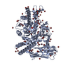 5fqeSC 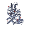 5fqfC 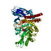 5fqgC  5fqhC S: Starting model for refinement C: citing same article ( |
|---|---|
| Similar structure data |


-Links
 PDBj
PDBj- Assembly
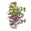
| Deposited unit | 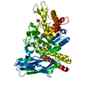
| ||||||||
|---|---|---|---|---|---|---|---|---|---|
| 1 |
| ||||||||
| Unit cell |
|
-Components
| #1: Protein | Mass: 70234.234 Da / Num. of mol.: 1 Source method: isolated from a genetically manipulated source Source: (gene. exp.) CLOSTRIDIUM PERFRINGENS (bacteria) / Production host: |
|---|---|
| #2: Sugar | ChemComp-SIZ /   Type: D-saccharide, beta linking / Mass: 257.189 Da / Num. of mol.: 1 Type: D-saccharide, beta linking / Mass: 257.189 Da / Num. of mol.: 1Source method: isolated from a genetically manipulated source Formula: C8H13F2NO6 |
| #3: Chemical | ChemComp-PO4 /   Mass: 94.971 Da / Num. of mol.: 1 / Source method: obtained synthetically / Formula: PO4 Mass: 94.971 Da / Num. of mol.: 1 / Source method: obtained synthetically / Formula: PO4 |
| #4: Water | ChemComp-HOH /  Mass: 18.015 Da / Num. of mol.: 415 / Source method: isolated from a natural source / Formula: H2O Mass: 18.015 Da / Num. of mol.: 415 / Source method: isolated from a natural source / Formula: H2O |
| Has protein modification | Y |
-Experimental details
-Experiment
| Experiment | Method: X-RAY DIFFRACTION / Number of used crystals: 1 |
|---|
- Sample preparation
| Crystal | Density Matthews: 1.94 Å3/Da / Density % sol: 36.72 % / Description: NONE |
|---|
-Data collection
| Diffraction | Mean temperature: 100 K |
|---|---|
| Diffraction source | Source: SYNCHROTRON / Site: CLSI  / Beamline: 08ID-1 / Wavelength: 0.984 / Beamline: 08ID-1 / Wavelength: 0.984 |
| Detector | Detector: CCD / Details: MIRRORS |
| Radiation | Protocol: SINGLE WAVELENGTH / Monochromatic (M) / Laue (L): M / Scattering type: x-ray |
| Radiation wavelength | Wavelength: 0.984 Å / Relative weight: 1 |
| Reflection | Resolution: 1.75→47.7 Å / Num. obs: 55369 / % possible obs: 100 % / Observed criterion σ(I): 2 / Redundancy: 12.6 % / Rmerge(I) obs: 0.11 / Net I/σ(I): 14.7 |
| Reflection shell | Resolution: 1.75→1.78 Å / Redundancy: 8.2 % / Rmerge(I) obs: 0.48 / Mean I/σ(I) obs: 2.6 / % possible all: 100 |
- Processing
| Software |
| ||||||||||||||||||||||||||||||||||||||||||||||||||||||||||||||||||||||||||||||||||||||||||||||||||||||||||||||||||||||||||||||||||||||||||||||||||||||||||||||||||||||||||||||||||||||
|---|---|---|---|---|---|---|---|---|---|---|---|---|---|---|---|---|---|---|---|---|---|---|---|---|---|---|---|---|---|---|---|---|---|---|---|---|---|---|---|---|---|---|---|---|---|---|---|---|---|---|---|---|---|---|---|---|---|---|---|---|---|---|---|---|---|---|---|---|---|---|---|---|---|---|---|---|---|---|---|---|---|---|---|---|---|---|---|---|---|---|---|---|---|---|---|---|---|---|---|---|---|---|---|---|---|---|---|---|---|---|---|---|---|---|---|---|---|---|---|---|---|---|---|---|---|---|---|---|---|---|---|---|---|---|---|---|---|---|---|---|---|---|---|---|---|---|---|---|---|---|---|---|---|---|---|---|---|---|---|---|---|---|---|---|---|---|---|---|---|---|---|---|---|---|---|---|---|---|---|---|---|---|---|
| Refinement | Method to determine structure: MOLECULAR REPLACEMENT Starting model: PDB ENTRY 5FQE Resolution: 1.75→95.31 Å / Cor.coef. Fo:Fc: 0.965 / Cor.coef. Fo:Fc free: 0.947 / SU B: 3.202 / SU ML: 0.101 / Cross valid method: THROUGHOUT / ESU R: 0.128 / ESU R Free: 0.122 / Stereochemistry target values: MAXIMUM LIKELIHOOD / Details: HYDROGENS HAVE BEEN ADDED IN THE RIDING POSITIONS.
| ||||||||||||||||||||||||||||||||||||||||||||||||||||||||||||||||||||||||||||||||||||||||||||||||||||||||||||||||||||||||||||||||||||||||||||||||||||||||||||||||||||||||||||||||||||||
| Solvent computation | Ion probe radii: 0.8 Å / Shrinkage radii: 0.8 Å / VDW probe radii: 1.2 Å / Solvent model: MASK | ||||||||||||||||||||||||||||||||||||||||||||||||||||||||||||||||||||||||||||||||||||||||||||||||||||||||||||||||||||||||||||||||||||||||||||||||||||||||||||||||||||||||||||||||||||||
| Displacement parameters | Biso mean: 35.234 Å2
| ||||||||||||||||||||||||||||||||||||||||||||||||||||||||||||||||||||||||||||||||||||||||||||||||||||||||||||||||||||||||||||||||||||||||||||||||||||||||||||||||||||||||||||||||||||||
| Refinement step | Cycle: LAST / Resolution: 1.75→95.31 Å
| ||||||||||||||||||||||||||||||||||||||||||||||||||||||||||||||||||||||||||||||||||||||||||||||||||||||||||||||||||||||||||||||||||||||||||||||||||||||||||||||||||||||||||||||||||||||
| Refine LS restraints |
|