 Movie
Movie Controller

[English] 日本語
 Yorodumi
- PDB-5fbp: CRYSTAL STRUCTURE OF THE NEUTRAL FORM OF FRUCTOSE-1,6-BISPHOSPHAT... -
+ Open data

- Basic information
| Entry | Database: PDB / ID: 5fbp | ||||||
|---|---|---|---|---|---|---|---|
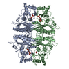
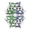
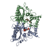
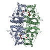
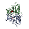
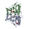
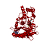
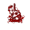
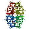
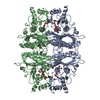
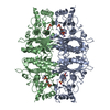
| Title | CRYSTAL STRUCTURE OF THE NEUTRAL FORM OF FRUCTOSE-1,6-BISPHOSPHATASE COMPLEXED WITH THE PRODUCT FRUCTOSE 6-PHOSPHATE AT 2.1-ANGSTROMS RESOLUTION | ||||||
 Components Components | FRUCTOSE 1,6-BISPHOSPHATASE | ||||||
 Keywords Keywords | HYDROLASE (PHOSPHORIC MONOESTER) | ||||||
| Function / homology |  Function and homology information Function and homology informationGluconeogenesis / fructose-bisphosphatase / fructose 1,6-bisphosphate 1-phosphatase activity / fructose 1,6-bisphosphate metabolic process / cellular response to magnesium ion / negative regulation of Ras protein signal transduction / fructose 6-phosphate metabolic process / fructose metabolic process / monosaccharide binding / negative regulation of glycolytic process ...Gluconeogenesis / fructose-bisphosphatase / fructose 1,6-bisphosphate 1-phosphatase activity / fructose 1,6-bisphosphate metabolic process / cellular response to magnesium ion / negative regulation of Ras protein signal transduction / fructose 6-phosphate metabolic process / fructose metabolic process / monosaccharide binding / negative regulation of glycolytic process / regulation of gluconeogenesis / AMP binding / gluconeogenesis / cellular response to xenobiotic stimulus / negative regulation of cell growth / RNA polymerase II-specific DNA-binding transcription factor binding / negative regulation of transcription by RNA polymerase II / metal ion binding / identical protein binding / nucleus / cytosol / cytoplasm Similarity search - Function | ||||||
| Biological species |  | ||||||
| Method |  X-RAY DIFFRACTION / Resolution: 2.1 Å X-RAY DIFFRACTION / Resolution: 2.1 Å | ||||||
 Authors Authors | Ke, H. / Liang, J.-Y. / Zhang, Y. / Lipscomb, W.N. | ||||||
 Citation Citation | Journal: Proc.Natl.Acad.Sci.USA / Year: 1991 Title: Crystal structure of the neutral form of fructose-1,6-bisphosphatase complexed with the product fructose 6-phosphate at 2.1-A resolution. Authors: Ke, H.M. / Zhang, Y.P. / Liang, J.Y. / Lipscomb, W.N. #1: Journal: Biochemistry / Year: 1991Title: Conformational Transition of Fructose-1,6-Bisphosphatase: Structure Comparison between the AMP Complex (T Form) and the Fructose 6-Phosphate Complex (R Form) Authors: Ke, H. / Liang, J.-Y. / Zhang, Y. / Lipscomb, W.N. | ||||||
| History |
|
- Structure visualization
| Structure viewer | Molecule: MolmilJmol/JSmol |
|---|
- Downloads & links
-Download
| PDBx/mmCIF format | 5fbp.cif.gz | 169.6 KB | Display | PDBx/mmCIF format |
|---|---|---|---|---|
| PDB format | pdb5fbp.ent.gz | 136.9 KB | Display | PDB format |
| PDBx/mmJSON format | 5fbp.json.gz | Tree view | PDBx/mmJSON format | |
| Others |  Other downloads Other downloads |
-Validation report
| Arichive directory | https://data.pdbj.org/pub/pdb/validation_reports/fb/5fbpftp://data.pdbj.org/pub/pdb/validation_reports/fb/5fbp | HTTPS FTP |
|---|
-Related structure data
| Similar structure data |
|---|


-Links
 PDBj
PDBj
- Assembly
| Deposited unit | 
| ||||||||
|---|---|---|---|---|---|---|---|---|---|
| 1 | 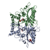
| ||||||||
| Unit cell |
|
-Components
| #1: Protein | Mass: 36503.004 Da / Num. of mol.: 2 Source method: isolated from a genetically manipulated source Source: (gene. exp.) #2: Sugar |   Type: D-saccharide, beta linking / Mass: 260.136 Da / Num. of mol.: 3 Type: D-saccharide, beta linking / Mass: 260.136 Da / Num. of mol.: 3Source method: isolated from a genetically manipulated source Formula: C6H13O9P #3: Water | ChemComp-HOH / |  Mass: 18.015 Da / Num. of mol.: 308 / Source method: isolated from a natural source / Formula: H2O Mass: 18.015 Da / Num. of mol.: 308 / Source method: isolated from a natural source / Formula: H2ONonpolymer details | THERE IS A MOLECULE OF F6P BOUND AT THE ACTIVE SITE IN BOTH CHAINS. THERE IS A MOLECULE OF F6P ...THERE IS A MOLECULE OF F6P BOUND AT THE ACTIVE SITE IN BOTH CHAINS. THERE IS A MOLECULE OF F6P BOUND AT THE AMP SITE IN CHAIN *A* BUT NOT IN CHAIN *B*. | |
|---|
-Experimental details
-Experiment
| Experiment | Method: X-RAY DIFFRACTION |
|---|
- Sample preparation
| Crystal | Density Matthews: 2.37 Å3/Da / Density % sol: 48.04 % | ||||||||||||||||||||||||||||||||||||
|---|---|---|---|---|---|---|---|---|---|---|---|---|---|---|---|---|---|---|---|---|---|---|---|---|---|---|---|---|---|---|---|---|---|---|---|---|---|
| Crystal grow | *PLUS pH: 7.4 / Method: microdialysis | ||||||||||||||||||||||||||||||||||||
| Components of the solutions | *PLUS
|
-Data collection
| Reflection | *PLUS Highest resolution: 2.1 Å / Num. obs: 38643 / % possible obs: 97 % / Num. measured all: 200580 |
|---|
- Processing
| Software | Name: X-PLOR / Classification: refinement | ||||||||||||||||||||||||||||||||||||||||||||||||||||||||||||
|---|---|---|---|---|---|---|---|---|---|---|---|---|---|---|---|---|---|---|---|---|---|---|---|---|---|---|---|---|---|---|---|---|---|---|---|---|---|---|---|---|---|---|---|---|---|---|---|---|---|---|---|---|---|---|---|---|---|---|---|---|---|
| Refinement | Rfactor Rwork: 0.177 / Highest resolution: 2.1 Å | ||||||||||||||||||||||||||||||||||||||||||||||||||||||||||||
| Refinement step | Cycle: LAST / Highest resolution: 2.1 Å
| ||||||||||||||||||||||||||||||||||||||||||||||||||||||||||||
| Refine LS restraints |
| ||||||||||||||||||||||||||||||||||||||||||||||||||||||||||||
| Software | *PLUS Name: X-PLOR / Classification: refinement | ||||||||||||||||||||||||||||||||||||||||||||||||||||||||||||
| Refinement | *PLUS Highest resolution: 2.1 Å / Lowest resolution: 10 Å / Num. reflection all: 35069 / Rfactor obs: 0.177 | ||||||||||||||||||||||||||||||||||||||||||||||||||||||||||||
| Solvent computation | *PLUS | ||||||||||||||||||||||||||||||||||||||||||||||||||||||||||||
| Displacement parameters | *PLUS Biso mean: 25.5 Å2 | ||||||||||||||||||||||||||||||||||||||||||||||||||||||||||||
| Refine LS restraints | *PLUS Type: x_angle_d / Dev ideal: 2.9 |
