 Movie
Movie Controller

[English] 日本語
 Yorodumi
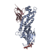
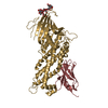
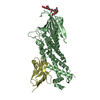
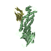
- PDB-5f9a: Blood group antigen binding adhesin BabA of Helicobacter pylori s... -
+ Open data

- Basic information
| Entry | Database: PDB / ID: 5f9a | ||||||||||||||||||
|---|---|---|---|---|---|---|---|---|---|---|---|---|---|---|---|---|---|---|---|
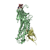
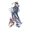
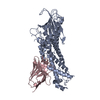
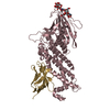
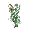
| Title | Blood group antigen binding adhesin BabA of Helicobacter pylori strain P436 in complex with blood group H Lewis b hexasaccharide | ||||||||||||||||||
 Components Components |
| ||||||||||||||||||
 Keywords Keywords | CELL ADHESION / Adhesin / Lectin / Nanobody / Complex | ||||||||||||||||||
| Function / homology |  Function and homology information Function and homology informationSabA, N-terminal extracellular adhesion domain / SabA N-terminal extracellular adhesion domain / Outer membrane protein, Helicobacter / Helicobacter outer membrane protein / Immunoglobulins / Immunoglobulin-like / Sandwich / Mainly Beta Similarity search - Domain/homology | ||||||||||||||||||
| Biological species |   Helicobacter pylori (bacteria) Helicobacter pylori (bacteria) | ||||||||||||||||||
| Method |  X-RAY DIFFRACTION / SYNCHROTRON / MOLECULAR REPLACEMENT / Resolution: 2.44 Å X-RAY DIFFRACTION / SYNCHROTRON / MOLECULAR REPLACEMENT / Resolution: 2.44 Å | ||||||||||||||||||
 Authors Authors | Moonens, K. / Gideonsson, P. / Subedi, S. / Romao, E. / Oscarson, S. / Muyldermans, S. / Boren, T. / Remaut, H. | ||||||||||||||||||
| Funding support |  Belgium, Belgium,  Sweden, 5items Sweden, 5items
| ||||||||||||||||||
 Citation Citation | Journal: Cell Host Microbe / Year: 2016 Title: Structural Insights into Polymorphic ABO Glycan Binding by Helicobacter pylori. Authors: Moonens, K. / Gideonsson, P. / Subedi, S. / Bugaytsova, J. / Romao, E. / Mendez, M. / Norden, J. / Fallah, M. / Rakhimova, L. / Shevtsova, A. / Lahmann, M. / Castaldo, G. / Brannstrom, K. / ...Authors: Moonens, K. / Gideonsson, P. / Subedi, S. / Bugaytsova, J. / Romao, E. / Mendez, M. / Norden, J. / Fallah, M. / Rakhimova, L. / Shevtsova, A. / Lahmann, M. / Castaldo, G. / Brannstrom, K. / Coppens, F. / Lo, A.W. / Ny, T. / Solnick, J.V. / Vandenbussche, G. / Oscarson, S. / Hammarstrom, L. / Arnqvist, A. / Berg, D.E. / Muyldermans, S. / Boren, T. / Remaut, H. | ||||||||||||||||||
| History |
|
- Structure visualization
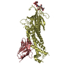
| Structure viewer | Molecule: MolmilJmol/JSmol |
|---|
- Downloads & links
-Download
| PDBx/mmCIF format | 5f9a.cif.gz | 220 KB | Display | PDBx/mmCIF format |
|---|---|---|---|---|
| PDB format | pdb5f9a.ent.gz | 174 KB | Display | PDB format |
| PDBx/mmJSON format | 5f9a.json.gz | Tree view | PDBx/mmJSON format | |
| Others |  Other downloads Other downloads |
-Validation report
| Arichive directory | https://data.pdbj.org/pub/pdb/validation_reports/f9/5f9aftp://data.pdbj.org/pub/pdb/validation_reports/f9/5f9a | HTTPS FTP |
|---|
-Related structure data
| Related structure data | 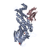 5f7kSC 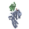 5f7lC 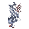 5f7mC  5f7nC 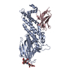 5f7wC  5f7yC  5f8qC 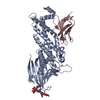 5f8rC 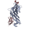 5f93C 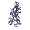 5f97C 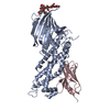 5f9dC S: Starting model for refinement C: citing same article ( |
|---|---|
| Similar structure data |


-Links
 PDBj
PDBj


- Assembly
| Deposited unit | 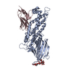
| ||||||||
|---|---|---|---|---|---|---|---|---|---|
| 1 |
| ||||||||
| Unit cell |
|
-Components
| #1: Protein | Mass: 48900.660 Da / Num. of mol.: 1 Source method: isolated from a genetically manipulated source Details: In this hybrid construct the insertion domain of the Peruvian strain P436 (residues 179 to 258, responsible for carbohydrate binding) was grafted into the framework of the generalist strain 17875 Source: (gene. exp.) Helicobacter pylori (bacteria) / Gene: babA2, babA / Production host: |
|---|---|
| #2: Antibody | Mass: 13260.856 Da / Num. of mol.: 1 Source method: isolated from a genetically manipulated source Source: (gene. exp.) |
| #3: Polysaccharide | alpha-L-fucopyranose-(1-2)-beta-D-galactopyranose-(1-3)-[alpha-L-fucopyranose-(1-4)]2-acetamido-2- ...alpha-L-fucopyranose-(1-2)-beta-D-galactopyranose-(1-3)-[alpha-L-fucopyranose-(1-4)]2-acetamido-2-deoxy-beta-D-glucopyranose-(1-3)-beta-D-galactopyranose-(1-4)-alpha-D-glucopyranose Source method: isolated from a genetically manipulated source |
| #4: Water | ChemComp-HOH /  Mass: 18.015 Da / Num. of mol.: 45 / Source method: isolated from a natural source / Formula: H2O Mass: 18.015 Da / Num. of mol.: 45 / Source method: isolated from a natural source / Formula: H2O |
| Has protein modification | Y |
-Experimental details
-Experiment
| Experiment | Method: X-RAY DIFFRACTION |
|---|
- Sample preparation
| Crystal | Density Matthews: 4.1 Å3/Da / Density % sol: 70.02 % |
|---|---|
| Crystal grow | Temperature: 293 K / Method: vapor diffusion, sitting drop / pH: 8 / Details: 1 M Sodium acetate, 100 mM Imidazole pH 8.0 |
-Data collection
| Diffraction | Mean temperature: 100 K |
|---|---|
| Diffraction source | Source: SYNCHROTRON / Site: Diamond  / Beamline: I04 / Wavelength: 0.98 Å / Beamline: I04 / Wavelength: 0.98 Å |
| Detector | Type: DECTRIS PILATUS 6M / Detector: PIXEL / Date: Jul 29, 2015 |
| Radiation | Protocol: SINGLE WAVELENGTH / Monochromatic (M) / Laue (L): M / Scattering type: x-ray |
| Radiation wavelength | Wavelength: 0.98 Å / Relative weight: 1 |
| Reflection | Resolution: 2.44→51.54 Å / Num. obs: 39582 / % possible obs: 100 % / Redundancy: 6.5 % / Rmerge(I) obs: 0.095 / Net I/σ(I): 8.8 |
| Reflection shell | Resolution: 2.44→2.57 Å / Redundancy: 6.4 % / Mean I/σ(I) obs: 1.7 / % possible all: 100 |
- Processing
| Software |
| ||||||||||||||||||||||||||||||||||||||||||||||||||||||||||||||||||||||||||||||||||||||||||||||||||||||||||||||||||||||||||||||||||||||||||||||||||||||||||||||||||||||||||||||||||||||
|---|---|---|---|---|---|---|---|---|---|---|---|---|---|---|---|---|---|---|---|---|---|---|---|---|---|---|---|---|---|---|---|---|---|---|---|---|---|---|---|---|---|---|---|---|---|---|---|---|---|---|---|---|---|---|---|---|---|---|---|---|---|---|---|---|---|---|---|---|---|---|---|---|---|---|---|---|---|---|---|---|---|---|---|---|---|---|---|---|---|---|---|---|---|---|---|---|---|---|---|---|---|---|---|---|---|---|---|---|---|---|---|---|---|---|---|---|---|---|---|---|---|---|---|---|---|---|---|---|---|---|---|---|---|---|---|---|---|---|---|---|---|---|---|---|---|---|---|---|---|---|---|---|---|---|---|---|---|---|---|---|---|---|---|---|---|---|---|---|---|---|---|---|---|---|---|---|---|---|---|---|---|---|---|
| Refinement | Method to determine structure: MOLECULAR REPLACEMENT Starting model: 5F7K Resolution: 2.44→51.54 Å / Cor.coef. Fo:Fc: 0.951 / Cor.coef. Fo:Fc free: 0.927 / SU B: 18.014 / SU ML: 0.18 / Cross valid method: THROUGHOUT / ESU R: 0.225 / ESU R Free: 0.212 / Stereochemistry target values: MAXIMUM LIKELIHOOD / Details: HYDROGENS HAVE BEEN ADDED IN THE RIDING POSITIONS
| ||||||||||||||||||||||||||||||||||||||||||||||||||||||||||||||||||||||||||||||||||||||||||||||||||||||||||||||||||||||||||||||||||||||||||||||||||||||||||||||||||||||||||||||||||||||
| Solvent computation | Ion probe radii: 0.8 Å / Shrinkage radii: 0.8 Å / VDW probe radii: 1.2 Å / Solvent model: MASK | ||||||||||||||||||||||||||||||||||||||||||||||||||||||||||||||||||||||||||||||||||||||||||||||||||||||||||||||||||||||||||||||||||||||||||||||||||||||||||||||||||||||||||||||||||||||
| Displacement parameters | Biso mean: 57.443 Å2
| ||||||||||||||||||||||||||||||||||||||||||||||||||||||||||||||||||||||||||||||||||||||||||||||||||||||||||||||||||||||||||||||||||||||||||||||||||||||||||||||||||||||||||||||||||||||
| Refinement step | Cycle: 1 / Resolution: 2.44→51.54 Å
| ||||||||||||||||||||||||||||||||||||||||||||||||||||||||||||||||||||||||||||||||||||||||||||||||||||||||||||||||||||||||||||||||||||||||||||||||||||||||||||||||||||||||||||||||||||||
| Refine LS restraints |
|