 Movie
Movie Controller

[English] 日本語
 Yorodumi
- PDB-5f8x: The crystal structure of human plasma kallikrein in complex with ... -
+ Open data

- Basic information
| Entry | Database: PDB / ID: 5f8x | |||||||||
|---|---|---|---|---|---|---|---|---|---|---|
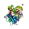
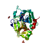
| Title | The crystal structure of human plasma kallikrein in complex with its peptide inhibitor pkalin-3 | |||||||||
 Components Components |
| |||||||||
 Keywords Keywords | HYDROLASE/HYDROLASE INHIBITOR / PEPTIDE INHIBITOR / HYDROLASE-HYDROLASE INHIBITOR COMPLEX | |||||||||
| Function / homology |  Function and homology information Function and homology informationplasma kallikrein / Factor XII activation / Defective SERPING1 causes hereditary angioedema / positive regulation of fibrinolysis / zymogen activation / plasminogen activation / Defective factor XII causes hereditary angioedema / Activation of Matrix Metalloproteinases / fibrinolysis / : ...plasma kallikrein / Factor XII activation / Defective SERPING1 causes hereditary angioedema / positive regulation of fibrinolysis / zymogen activation / plasminogen activation / Defective factor XII causes hereditary angioedema / Activation of Matrix Metalloproteinases / fibrinolysis / : / serine-type peptidase activity / blood coagulation / serine-type endopeptidase activity / proteolysis / : / extracellular exosome / extracellular region / plasma membrane Similarity search - Function | |||||||||
| Biological species |  Homo sapiens (human) Homo sapiens (human)synthetic construct (others) | |||||||||
| Method |  X-RAY DIFFRACTION / SYNCHROTRON / MOLECULAR REPLACEMENT / Resolution: 1.55 Å X-RAY DIFFRACTION / SYNCHROTRON / MOLECULAR REPLACEMENT / Resolution: 1.55 Å | |||||||||
 Authors Authors | Xu, M. / Jiang, L. / Xu, P. / Luo, Z. / Andreasen, P. / Huang, M. | |||||||||
 Citation Citation | Journal: To Be Published Title: The Crystal Structure Of Human Plasma Kallikrein In Complex With Its Peptide Inhibitor Pkalin-3 Authors: Xu, M. / Jiang, L. / Xu, P. / Luo, Z. / Andreasen, P. / Huang, M. | |||||||||
| History |
|
- Structure visualization
| Structure viewer | Molecule: MolmilJmol/JSmol |
|---|
- Downloads & links
-Download
| PDBx/mmCIF format | 5f8x.cif.gz | 69.7 KB | Display | PDBx/mmCIF format |
|---|---|---|---|---|
| PDB format | pdb5f8x.ent.gz | 49.6 KB | Display | PDB format |
| PDBx/mmJSON format | 5f8x.json.gz | Tree view | PDBx/mmJSON format | |
| Others |  Other downloads Other downloads |
-Validation report
| Arichive directory | https://data.pdbj.org/pub/pdb/validation_reports/f8/5f8xftp://data.pdbj.org/pub/pdb/validation_reports/f8/5f8x | HTTPS FTP |
|---|
-Related structure data
| Related structure data |  2anyS S: Starting model for refinement |
|---|---|
| Similar structure data |


-Links
 PDBj
PDBj


- Assembly
| Deposited unit | 
| ||||||||
|---|---|---|---|---|---|---|---|---|---|
| 1 |
| ||||||||
| Unit cell |
|
-Components
| #1: Protein | Mass: 26913.578 Da / Num. of mol.: 1 / Fragment: UNP RESIDUES 391-629 / Mutation: C122S Source method: isolated from a genetically manipulated source Source: (gene. exp.) Homo sapiens (human) / Gene: KLKB1, KLK3 / Production host:  Komagataella pastoris (fungus) / References: UniProt: P03952, plasma kallikrein Komagataella pastoris (fungus) / References: UniProt: P03952, plasma kallikrein | ||||||
|---|---|---|---|---|---|---|---|
| #2: Protein/peptide | Mass: 1138.384 Da / Num. of mol.: 1 / Source method: obtained synthetically / Source: (synth.) synthetic construct (others) | ||||||
| #3: Chemical | ChemComp-SO4 /   Mass: 96.063 Da / Num. of mol.: 8 / Source method: obtained synthetically / Formula: SO4 Mass: 96.063 Da / Num. of mol.: 8 / Source method: obtained synthetically / Formula: SO4#4: Chemical | ChemComp-MRZ / |   Mass: 127.188 Da / Num. of mol.: 1 / Source method: obtained synthetically / Formula: C6H13N3 Mass: 127.188 Da / Num. of mol.: 1 / Source method: obtained synthetically / Formula: C6H13N3#5: Water | ChemComp-HOH / |  Mass: 18.015 Da / Num. of mol.: 195 / Source method: isolated from a natural source / Formula: H2O Mass: 18.015 Da / Num. of mol.: 195 / Source method: isolated from a natural source / Formula: H2OHas protein modification | Y | |
-Experimental details
-Experiment
| Experiment | Method: X-RAY DIFFRACTION / Number of used crystals: 1 |
|---|
- Sample preparation
| Crystal | Density Matthews: 2.41 Å3/Da / Density % sol: 49 % |
|---|---|
| Crystal grow | Temperature: 298 K / Method: vapor diffusion, sitting drop / pH: 8.5 Details: 23% PEG 3350, 0.1M TRIS-HCL, PH 8.5, 20MM (NH4)2SO4, VAPOR DIFFUSION, SITTING DROP, TEMPERATURE 298K |
-Data collection
| Diffraction | Mean temperature: 100 K |
|---|---|
| Diffraction source | Source: SYNCHROTRON / Site: SSRF  / Beamline: BL17U / Wavelength: 0.97 Å / Beamline: BL17U / Wavelength: 0.97 Å |
| Detector | Type: ADSC QUANTUM 315r / Detector: CCD / Date: Jul 11, 2014 |
| Radiation | Protocol: SINGLE WAVELENGTH / Monochromatic (M) / Laue (L): M / Scattering type: x-ray |
| Radiation wavelength | Wavelength: 0.97 Å / Relative weight: 1 |
| Reflection | Resolution: 1.55→50 Å / Num. obs: 38722 / % possible obs: 100 % / Redundancy: 5.5 % / Net I/σ(I): 26.2 |
- Processing
| Software |
| ||||||||||||||||||||||||||||||||||||||||||||||||||||||||||||||||||||||||||||||||||||||||||||||||||||||||||||||||||||||||||||||||||||||||||||||||||||||||||||||||||||||||||||||||||||||
|---|---|---|---|---|---|---|---|---|---|---|---|---|---|---|---|---|---|---|---|---|---|---|---|---|---|---|---|---|---|---|---|---|---|---|---|---|---|---|---|---|---|---|---|---|---|---|---|---|---|---|---|---|---|---|---|---|---|---|---|---|---|---|---|---|---|---|---|---|---|---|---|---|---|---|---|---|---|---|---|---|---|---|---|---|---|---|---|---|---|---|---|---|---|---|---|---|---|---|---|---|---|---|---|---|---|---|---|---|---|---|---|---|---|---|---|---|---|---|---|---|---|---|---|---|---|---|---|---|---|---|---|---|---|---|---|---|---|---|---|---|---|---|---|---|---|---|---|---|---|---|---|---|---|---|---|---|---|---|---|---|---|---|---|---|---|---|---|---|---|---|---|---|---|---|---|---|---|---|---|---|---|---|---|
| Refinement | Method to determine structure: MOLECULAR REPLACEMENT Starting model: 2ANY Resolution: 1.55→50 Å / Cor.coef. Fo:Fc: 0.944 / Cor.coef. Fo:Fc free: 0.932 / SU B: 1.729 / SU ML: 0.064 / Cross valid method: THROUGHOUT / ESU R: 0.099 / ESU R Free: 0.095 / Stereochemistry target values: MAXIMUM LIKELIHOOD / Details: HYDROGENS HAVE BEEN ADDED IN THE RIDING POSITIONS
| ||||||||||||||||||||||||||||||||||||||||||||||||||||||||||||||||||||||||||||||||||||||||||||||||||||||||||||||||||||||||||||||||||||||||||||||||||||||||||||||||||||||||||||||||||||||
| Solvent computation | Ion probe radii: 0.8 Å / Shrinkage radii: 0.8 Å / VDW probe radii: 1.2 Å / Solvent model: MASK | ||||||||||||||||||||||||||||||||||||||||||||||||||||||||||||||||||||||||||||||||||||||||||||||||||||||||||||||||||||||||||||||||||||||||||||||||||||||||||||||||||||||||||||||||||||||
| Displacement parameters | Biso mean: 21.91 Å2
| ||||||||||||||||||||||||||||||||||||||||||||||||||||||||||||||||||||||||||||||||||||||||||||||||||||||||||||||||||||||||||||||||||||||||||||||||||||||||||||||||||||||||||||||||||||||
| Refinement step | Cycle: LAST / Resolution: 1.55→50 Å
| ||||||||||||||||||||||||||||||||||||||||||||||||||||||||||||||||||||||||||||||||||||||||||||||||||||||||||||||||||||||||||||||||||||||||||||||||||||||||||||||||||||||||||||||||||||||
| Refine LS restraints |
|