 Movie
Movie Controller

[English] 日本語
 Yorodumi
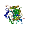
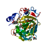
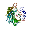
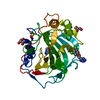
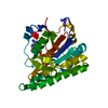
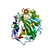
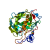
- PDB-5f31: Crystal structure of membrane associated PatA from Mycobacterium ... -
+ Open data

- Basic information
| Entry | Database: PDB / ID: 5f31 | ||||||
|---|---|---|---|---|---|---|---|
| Title | Crystal structure of membrane associated PatA from Mycobacterium smegmatis in complex with palmitate - P 42 21 2 space group | ||||||
 Components Components | Phosphatidylinositol mannoside acyltransferase | ||||||
 Keywords Keywords | TRANSFERASE / acyltransferase / glycolipid biosynthesis | ||||||
| Function / homology |  Function and homology information Function and homology informationphosphatidylinositol dimannoside acyltransferase / glycolipid biosynthetic process / phosphatidylinositol metabolic process / phospholipid biosynthetic process / acyltransferase activity / plasma membrane Similarity search - Function | ||||||
| Biological species |  Mycobacterium smegmatis str. MC2 155 (bacteria) Mycobacterium smegmatis str. MC2 155 (bacteria) | ||||||
| Method |  X-RAY DIFFRACTION / SYNCHROTRON / MOLECULAR REPLACEMENT / Resolution: 2.43 Å X-RAY DIFFRACTION / SYNCHROTRON / MOLECULAR REPLACEMENT / Resolution: 2.43 Å | ||||||
 Authors Authors | Albesa-Jove, D. / Svetlikova, Z. / Carreras-Gonzalez, A. / Tersa, M. / Sancho-Vaello, E. / Cifuente, J.O. / Mikusova, K. / Guerin, M.E. | ||||||
 Citation Citation | Journal: Nat Commun / Year: 2016 Title: Structural basis for selective recognition of acyl chains by the membrane-associated acyltransferase PatA. Authors: Albesa-Jove, D. / Svetlikova, Z. / Tersa, M. / Sancho-Vaello, E. / Carreras-Gonzalez, A. / Bonnet, P. / Arrasate, P. / Eguskiza, A. / Angala, S.K. / Cifuente, J.O. / Kordulakova, J. / ...Authors: Albesa-Jove, D. / Svetlikova, Z. / Tersa, M. / Sancho-Vaello, E. / Carreras-Gonzalez, A. / Bonnet, P. / Arrasate, P. / Eguskiza, A. / Angala, S.K. / Cifuente, J.O. / Kordulakova, J. / Jackson, M. / Mikusova, K. / Guerin, M.E. | ||||||
| History |
|
- Structure visualization
| Structure viewer | Molecule: MolmilJmol/JSmol |
|---|
- Downloads & links
-Download
| PDBx/mmCIF format | 5f31.cif.gz | 68.6 KB | Display | PDBx/mmCIF format |
|---|---|---|---|---|
| PDB format | pdb5f31.ent.gz | 48.5 KB | Display | PDB format |
| PDBx/mmJSON format | 5f31.json.gz | Tree view | PDBx/mmJSON format | |
| Others |  Other downloads Other downloads |
-Validation report
| Arichive directory | https://data.pdbj.org/pub/pdb/validation_reports/f3/5f31ftp://data.pdbj.org/pub/pdb/validation_reports/f3/5f31 | HTTPS FTP |
|---|
-Related structure data


-Links
 PDBj
PDBj


- Assembly
| Deposited unit | 
| ||||||||||||||||||
|---|---|---|---|---|---|---|---|---|---|---|---|---|---|---|---|---|---|---|---|
| 1 |
| ||||||||||||||||||
| Unit cell |
| ||||||||||||||||||
| Components on special symmetry positions |
|
-Components
| #1: Protein | Mass: 34298.754 Da / Num. of mol.: 1 Source method: isolated from a genetically manipulated source Source: (gene. exp.) Mycobacterium smegmatis str. MC2 155 (bacteria)Gene: MSMEG_2934, MSMEI_2860 / Plasmid: pJAM2::patA Production host: Mycobacterium smegmatis str. MC2 155 (bacteria)References: UniProt: A0QWG5, Transferases; Acyltransferases; Transferring groups other than aminoacyl groups |
|---|---|
| #2: Chemical | ChemComp-PLM /   Mass: 256.424 Da / Num. of mol.: 1 / Source method: obtained synthetically / Formula: C16H32O2 Mass: 256.424 Da / Num. of mol.: 1 / Source method: obtained synthetically / Formula: C16H32O2 |
| #3: Chemical | ChemComp-EOH /   Mass: 46.068 Da / Num. of mol.: 1 / Source method: obtained synthetically / Formula: C2H6O Mass: 46.068 Da / Num. of mol.: 1 / Source method: obtained synthetically / Formula: C2H6O |
| #4: Chemical | ChemComp-NA /   Mass: 22.990 Da / Num. of mol.: 1 / Source method: obtained synthetically / Formula: Na Mass: 22.990 Da / Num. of mol.: 1 / Source method: obtained synthetically / Formula: Na |
| #5: Water | ChemComp-HOH /  Mass: 18.015 Da / Num. of mol.: 87 / Source method: isolated from a natural source / Formula: H2O Mass: 18.015 Da / Num. of mol.: 87 / Source method: isolated from a natural source / Formula: H2O |
-Experimental details
-Experiment
| Experiment | Method: X-RAY DIFFRACTION |
|---|
- Sample preparation
| Crystal | Density Matthews: 2.67 Å3/Da / Density % sol: 53.99 % |
|---|---|
| Crystal grow | Temperature: 291 K / Method: vapor diffusion, sitting drop / pH: 8.5 / Details: 100 mM Tris-HCl pH 8.5 and 20% ethanol |
-Data collection
| Diffraction | Mean temperature: 100 K |
|---|---|
| Diffraction source | Source: SYNCHROTRON / Site: SLS  / Beamline: X10SA / Wavelength: 0.99996 Å / Beamline: X10SA / Wavelength: 0.99996 Å |
| Detector | Type: DECTRIS PILATUS 6M / Detector: PIXEL / Date: Dec 19, 2011 |
| Radiation | Protocol: SINGLE WAVELENGTH / Monochromatic (M) / Laue (L): M / Scattering type: x-ray |
| Radiation wavelength | Wavelength: 0.99996 Å / Relative weight: 1 |
| Reflection | Resolution: 2.43→40.19 Å / Num. obs: 14652 / % possible obs: 100 % / Redundancy: 10.5 % / Rmerge(I) obs: 0.122 / Net I/σ(I): 4.6 |
| Reflection shell | Highest resolution: 2.43 Å / Redundancy: 10.9 % / Rmerge(I) obs: 0.669 / Mean I/σ(I) obs: 1.1 / % possible all: 100 |
- Processing
| Software |
| |||||||||||||||||||||||||||||||||||||||||||||||||
|---|---|---|---|---|---|---|---|---|---|---|---|---|---|---|---|---|---|---|---|---|---|---|---|---|---|---|---|---|---|---|---|---|---|---|---|---|---|---|---|---|---|---|---|---|---|---|---|---|---|---|
| Refinement | Method to determine structure: MOLECULAR REPLACEMENT / Resolution: 2.43→40.187 Å / SU ML: 0.26 / Cross valid method: FREE R-VALUE / σ(F): 1.35 / Phase error: 27.71 / Stereochemistry target values: ML
| |||||||||||||||||||||||||||||||||||||||||||||||||
| Solvent computation | Shrinkage radii: 0.9 Å / VDW probe radii: 1.11 Å / Solvent model: FLAT BULK SOLVENT MODEL | |||||||||||||||||||||||||||||||||||||||||||||||||
| Refinement step | Cycle: LAST / Resolution: 2.43→40.187 Å
| |||||||||||||||||||||||||||||||||||||||||||||||||
| Refine LS restraints |
| |||||||||||||||||||||||||||||||||||||||||||||||||
| LS refinement shell |
|
