 Movie
Movie Controller


+ Open data

- Basic information
| Entry | Database: PDB / ID: 5ez2 | ||||||
|---|---|---|---|---|---|---|---|
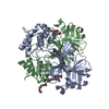
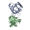
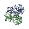
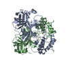
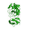
| Title | Sandercyanin Fluorescent Protein (SFP) | ||||||
 Components Components | Sandercyanin Fluorescent Protein | ||||||
 Keywords Keywords | FLUORESCENT PROTEIN / Sandercyanin / red-fluorescent protein / lipocalin / biliverdin / photo-stability | ||||||
| Function / homology |  Function and homology information Function and homology informationpigment binding / response to reactive oxygen species / lipid metabolic process / lipid binding / extracellular region / cytoplasm Similarity search - Function | ||||||
| Biological species |  Sander vitreus (walleye) Sander vitreus (walleye) | ||||||
| Method |  X-RAY DIFFRACTION / SYNCHROTRON / MOLECULAR REPLACEMENT / Resolution: 1.849 Å X-RAY DIFFRACTION / SYNCHROTRON / MOLECULAR REPLACEMENT / Resolution: 1.849 Å | ||||||
 Authors Authors | Ghosh, S. / Ramaswamy, S. | ||||||
 Citation Citation | Journal: Proc.Natl.Acad.Sci.USA / Year: 2016 Title: Blue protein with red fluorescence Authors: Ghosh, S. / Yu, C.L. / Ferraro, D.J. / Sudha, S. / Pal, S.K. / Schaefer, W.F. / Gibson, D.T. / Ramaswamy, S. | ||||||
| History |
|
- Structure visualization
| Structure viewer | Molecule: MolmilJmol/JSmol |
|---|
- Downloads & links
-Download
| PDBx/mmCIF format | 5ez2.cif.gz | 91.3 KB | Display | PDBx/mmCIF format |
|---|---|---|---|---|
| PDB format | pdb5ez2.ent.gz | 68.8 KB | Display | PDB format |
| PDBx/mmJSON format | 5ez2.json.gz | Tree view | PDBx/mmJSON format | |
| Others |  Other downloads Other downloads |
-Validation report
| Arichive directory | https://data.pdbj.org/pub/pdb/validation_reports/ez/5ez2ftp://data.pdbj.org/pub/pdb/validation_reports/ez/5ez2 | HTTPS FTP |
|---|
-Related structure data
| Related structure data |  5f1eC  5f6zSC C: citing same article ( S: Starting model for refinement |
|---|---|
| Similar structure data |

-Links
 PDBj
PDBj

- Assembly
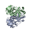
| Deposited unit | 
| |||||||||
|---|---|---|---|---|---|---|---|---|---|---|
| 1 | 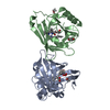
| |||||||||
| Unit cell |
| |||||||||
| Components on special symmetry positions |
|
-Components
| #1: Protein | Mass: 20159.748 Da / Num. of mol.: 2 Source method: isolated from a genetically manipulated source Source: (gene. exp.) Sander vitreus (walleye) / Tissue: Mucus / Cell line: Mucosa / Plasmid: pET21aDetails (production host): Ampicillin resistant plasmid, gene is cloned between NdeI and HindIII Production host:  #2: Chemical |   Mass: 582.646 Da / Num. of mol.: 2 Mass: 582.646 Da / Num. of mol.: 2Source method: isolated from a genetically manipulated source Formula: C33H34N4O6 #3: Chemical | ChemComp-EDO /   Mass: 62.068 Da / Num. of mol.: 5 / Source method: obtained synthetically / Formula: C2H6O2 Mass: 62.068 Da / Num. of mol.: 5 / Source method: obtained synthetically / Formula: C2H6O2#4: Water | ChemComp-HOH / |  Mass: 18.015 Da / Num. of mol.: 313 / Source method: isolated from a natural source / Formula: H2O Mass: 18.015 Da / Num. of mol.: 313 / Source method: isolated from a natural source / Formula: H2OHas protein modification | Y | |
|---|
-Experimental details
-Experiment
| Experiment | Method: X-RAY DIFFRACTION / Number of used crystals: 1 |
|---|
- Sample preparation
| Crystal | Density Matthews: 4.12 Å3/Da / Density % sol: 70.16 % |
|---|---|
| Crystal grow | Temperature: 277 K / Method: vapor diffusion, hanging drop / pH: 5.5 / Details: 0.1M Bis-Tris, 1M NaCl, PEG 400 / Temp details: Crystals were grown in a cold incubator |
-Data collection
| Diffraction | Mean temperature: 100 K |
|---|---|
| Diffraction source | Source: SYNCHROTRON / Site: ESRF  / Beamline: BM14 / Wavelength: 0.987 Å / Beamline: BM14 / Wavelength: 0.987 Å |
| Detector | Type: MAR CCD 130 mm / Detector: CCD / Date: Oct 10, 2013 |
| Radiation | Protocol: SINGLE WAVELENGTH / Monochromatic (M) / Laue (L): M / Scattering type: x-ray |
| Radiation wavelength | Wavelength: 0.987 Å / Relative weight: 1 |
| Reflection | Resolution: 1.849→35.99 Å / Num. obs: 53181 / % possible obs: 98.47 % / Redundancy: 4 % / Net I/σ(I): 15.48 |
| Reflection shell | Resolution: 1.849→1.915 Å |
- Processing
| Software |
| |||||||||||||||||||||||||||||||||||||||||||||||||||||||||||||||||||||||||||||||||||||||||||||||||||||||||
|---|---|---|---|---|---|---|---|---|---|---|---|---|---|---|---|---|---|---|---|---|---|---|---|---|---|---|---|---|---|---|---|---|---|---|---|---|---|---|---|---|---|---|---|---|---|---|---|---|---|---|---|---|---|---|---|---|---|---|---|---|---|---|---|---|---|---|---|---|---|---|---|---|---|---|---|---|---|---|---|---|---|---|---|---|---|---|---|---|---|---|---|---|---|---|---|---|---|---|---|---|---|---|---|---|---|---|
| Refinement | Method to determine structure: MOLECULAR REPLACEMENT Starting model: 5F6Z Resolution: 1.849→35.99 Å / SU ML: 0.19 / Cross valid method: FREE R-VALUE / σ(F): 1.34 / Phase error: 21.02 / Stereochemistry target values: ML
| |||||||||||||||||||||||||||||||||||||||||||||||||||||||||||||||||||||||||||||||||||||||||||||||||||||||||
| Solvent computation | Shrinkage radii: 0.9 Å / VDW probe radii: 1.11 Å / Solvent model: FLAT BULK SOLVENT MODEL | |||||||||||||||||||||||||||||||||||||||||||||||||||||||||||||||||||||||||||||||||||||||||||||||||||||||||
| Refinement step | Cycle: LAST / Resolution: 1.849→35.99 Å
| |||||||||||||||||||||||||||||||||||||||||||||||||||||||||||||||||||||||||||||||||||||||||||||||||||||||||
| Refine LS restraints |
| |||||||||||||||||||||||||||||||||||||||||||||||||||||||||||||||||||||||||||||||||||||||||||||||||||||||||
| LS refinement shell |
|
