 Movie
Movie Controller

[English] 日本語
 Yorodumi
- PDB-5epy: Crystal structure of HCV NS3/4A protease A156T variant in complex... -
+ Open data

- Basic information
| Entry | Database: PDB / ID: 5epy | |||||||||
|---|---|---|---|---|---|---|---|---|---|---|
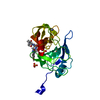
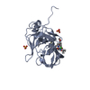
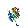
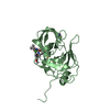
| Title | Crystal structure of HCV NS3/4A protease A156T variant in complex with 5172-mcP1P3 (MK-5172 P1-P3 macrocyclic analogue) | |||||||||
 Components Components | NS3 protease | |||||||||
 Keywords Keywords | HYDROLASE / grazoprevir analogue / macrocycle / drug resistance / HCV protease inhibitor / MK-5172 | |||||||||
| Function / homology |  Function and homology information Function and homology informationsymbiont-mediated transformation of host cell / host cell membrane / serine-type peptidase activity / viral capsid / host cell / RNA helicase activity / symbiont entry into host cell / virion attachment to host cell / virion membrane / proteolysis / metal ion binding Similarity search - Function | |||||||||
| Biological species |  Hepatitis C virus Hepatitis C virus | |||||||||
| Method |  X-RAY DIFFRACTION / SYNCHROTRON / MOLECULAR REPLACEMENT / molecular replacement / Resolution: 2.3 Å X-RAY DIFFRACTION / SYNCHROTRON / MOLECULAR REPLACEMENT / molecular replacement / Resolution: 2.3 Å | |||||||||
 Authors Authors | Soumana, D.I. / Yilmaz, N.K. / Ali, A. / Prachanronarong, K.L. / Aydin, C. / Schiffer, C.A. | |||||||||
| Funding support |  United States, 2items United States, 2items
| |||||||||
 Citation Citation | Journal: Acs Chem.Biol. / Year: 2016 Title: Structural and Thermodynamic Effects of Macrocyclization in HCV NS3/4A Inhibitor MK-5172. Authors: Soumana, D.I. / Kurt Yilmaz, N. / Prachanronarong, K.L. / Aydin, C. / Ali, A. / Schiffer, C.A. #1: Journal: ACS Chem. Biol. / Year: 2013 Title: Evaluating the role of macrocycles in the susceptibility of hepatitis C virus NS3/4A protease inhibitors to drug resistance. Authors: Ali, A. / Aydin, C. / Gildemeister, R. / Romano, K.P. / Cao, H. / Ozen, A. / Soumana, D. / Newton, A. / Petropoulos, C.J. / Huang, W. / Schiffer, C.A. | |||||||||
| History |
|
- Structure visualization
| Structure viewer | Molecule: MolmilJmol/JSmol |
|---|
- Downloads & links
-Download
| PDBx/mmCIF format | 5epy.cif.gz | 84.3 KB | Display | PDBx/mmCIF format |
|---|---|---|---|---|
| PDB format | pdb5epy.ent.gz | 61.6 KB | Display | PDB format |
| PDBx/mmJSON format | 5epy.json.gz | Tree view | PDBx/mmJSON format | |
| Others |  Other downloads Other downloads |
-Validation report
| Arichive directory | https://data.pdbj.org/pub/pdb/validation_reports/ep/5epyftp://data.pdbj.org/pub/pdb/validation_reports/ep/5epy | HTTPS FTP |
|---|
-Related structure data
| Related structure data |  5epnC 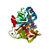 5eqqC  5etxC  3m5mS C: citing same article ( S: Starting model for refinement |
|---|---|
| Similar structure data |


-Links
 PDBj
PDBj
- Assembly
| Deposited unit | 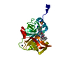
| ||||||||
|---|---|---|---|---|---|---|---|---|---|
| 1 |
| ||||||||
| Unit cell |
|
-Components
| #1: Protein | Mass: 21031.838 Da / Num. of mol.: 1 / Mutation: A156T Source method: isolated from a genetically manipulated source Source: (gene. exp.) Hepatitis C virus / Plasmid: pET28-a / Production host:  |
|---|---|
| #2: Chemical | ChemComp-ZN /   Mass: 65.409 Da / Num. of mol.: 1 / Source method: obtained synthetically / Formula: Zn Mass: 65.409 Da / Num. of mol.: 1 / Source method: obtained synthetically / Formula: Zn |
| #3: Chemical | ChemComp-SO4 /   Mass: 96.063 Da / Num. of mol.: 1 / Source method: obtained synthetically / Formula: SO4 Mass: 96.063 Da / Num. of mol.: 1 / Source method: obtained synthetically / Formula: SO4 |
| #4: Chemical | ChemComp-5R2 /   Mass: 754.893 Da / Num. of mol.: 1 / Source method: obtained synthetically / Formula: C37H50N6O9S Mass: 754.893 Da / Num. of mol.: 1 / Source method: obtained synthetically / Formula: C37H50N6O9S |
| #5: Water | ChemComp-HOH /  Mass: 18.015 Da / Num. of mol.: 78 / Source method: isolated from a natural source / Formula: H2O Mass: 18.015 Da / Num. of mol.: 78 / Source method: isolated from a natural source / Formula: H2O |
-Experimental details
-Experiment
| Experiment | Method: X-RAY DIFFRACTION / Number of used crystals: 1 |
|---|
- Sample preparation
| Crystal | Density Matthews: 2.3 Å3/Da / Density % sol: 46.42 % |
|---|---|
| Crystal grow | Temperature: 298 K / Method: vapor diffusion, hanging drop / pH: 6.5 Details: 100mM MES buffer pH 6.5, 4% (w/v) ammonium sulfate, 20-26% PEG 3350 |
-Data collection
| Diffraction | Mean temperature: 100 K | ||||||||||||||||||||||||||||||||||||||||||||||||||||||||||||||||||
|---|---|---|---|---|---|---|---|---|---|---|---|---|---|---|---|---|---|---|---|---|---|---|---|---|---|---|---|---|---|---|---|---|---|---|---|---|---|---|---|---|---|---|---|---|---|---|---|---|---|---|---|---|---|---|---|---|---|---|---|---|---|---|---|---|---|---|---|
| Diffraction source | Source: SYNCHROTRON / Site: APS / Beamline: 21-ID-G / Wavelength: 0.97857 Å | ||||||||||||||||||||||||||||||||||||||||||||||||||||||||||||||||||
| Detector | Type: MARMOSAIC 300 mm CCD / Detector: CCD / Date: Mar 14, 2013 | ||||||||||||||||||||||||||||||||||||||||||||||||||||||||||||||||||
| Radiation | Protocol: SINGLE WAVELENGTH / Monochromatic (M) / Laue (L): M / Scattering type: x-ray | ||||||||||||||||||||||||||||||||||||||||||||||||||||||||||||||||||
| Radiation wavelength | Wavelength: 0.97857 Å / Relative weight: 1 | ||||||||||||||||||||||||||||||||||||||||||||||||||||||||||||||||||
| Reflection | Resolution: 2.3→35 Å / Num. obs: 9070 / % possible obs: 100 % / Redundancy: 4.4 % / Rmerge(I) obs: 0.068 / Χ2: 1.075 / Net I/av σ(I): 22.401 / Net I/σ(I): 9.1 / Num. measured all: 39511 | ||||||||||||||||||||||||||||||||||||||||||||||||||||||||||||||||||
| Reflection shell | Diffraction-ID: 1 / Rejects: _
|
-Phasing
| Phasing | Method: molecular replacement |
|---|
- Processing
| Software |
| ||||||||||||||||||||
|---|---|---|---|---|---|---|---|---|---|---|---|---|---|---|---|---|---|---|---|---|---|
| Refinement | Method to determine structure: MOLECULAR REPLACEMENT Starting model: 3m5m Resolution: 2.3→35 Å / Cross valid method: FREE R-VALUE
| ||||||||||||||||||||
| Solvent computation | Shrinkage radii: 0.9 Å / VDW probe radii: 1.11 Å | ||||||||||||||||||||
| Displacement parameters | Biso max: 71.59 Å2 / Biso mean: 20.3824 Å2 / Biso min: 7.63 Å2 | ||||||||||||||||||||
| Refinement step | Cycle: LAST / Resolution: 2.3→35 Å
|
