 Movie
Movie Controller


+ Open data

- Basic information
| Entry | Database: PDB / ID: 5.0E+84 | ||||||
|---|---|---|---|---|---|---|---|
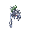
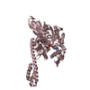
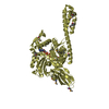
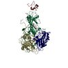
| Title | ATP-bound state of BiP | ||||||
 Components Components | 78 kDa glucose-regulated protein | ||||||
 Keywords Keywords | CHAPERONE / molecular chaperones / Hsp70 / BiP / Protein Folding / endoplasmic reticulum / allosteric coupling | ||||||
| Function / homology |  Function and homology information Function and homology informationregulation of ATF6-mediated unfolded protein response / regulation of PERK-mediated unfolded protein response / regulation of protein folding in endoplasmic reticulum / cerebellum structural organization / ATF6 (ATF6-alpha) activates chaperones / ATF6B (ATF6-beta) activates chaperones / maintenance of protein localization in endoplasmic reticulum / IRE1alpha activates chaperones / ATF6 (ATF6-alpha) activates chaperone genes / endoplasmic reticulum chaperone complex ...regulation of ATF6-mediated unfolded protein response / regulation of PERK-mediated unfolded protein response / regulation of protein folding in endoplasmic reticulum / cerebellum structural organization / ATF6 (ATF6-alpha) activates chaperones / ATF6B (ATF6-beta) activates chaperones / maintenance of protein localization in endoplasmic reticulum / IRE1alpha activates chaperones / ATF6 (ATF6-alpha) activates chaperone genes / endoplasmic reticulum chaperone complex / negative regulation of IRE1-mediated unfolded protein response / regulation of IRE1-mediated unfolded protein response / PERK regulates gene expression / protein folding in endoplasmic reticulum / cerebellar Purkinje cell layer development / misfolded protein binding / post-translational protein targeting to membrane, translocation / Modulation of host responses by IFN-stimulated genes / ER overload response / IRE1-mediated unfolded protein response / negative regulation of PERK-mediated unfolded protein response / endoplasmic reticulum-Golgi intermediate compartment / non-chaperonin molecular chaperone ATPase / intracellular membrane-bounded organelle / Regulation of HSF1-mediated heat shock response / protein serine/threonine kinase inhibitor activity / negative regulation of protein-containing complex assembly / cellular response to glucose starvation / cellular response to interleukin-4 / endoplasmic reticulum unfolded protein response / heat shock protein binding / ERAD pathway / protein folding chaperone / substantia nigra development / response to endoplasmic reticulum stress / positive regulation of protein ubiquitination / Antigen Presentation: Folding, assembly and peptide loading of class I MHC / ATP-dependent protein folding chaperone / protein sequestering activity / negative regulation of transforming growth factor beta receptor signaling pathway / : / melanosome / Platelet degranulation / protein refolding / protein-folding chaperone binding / ribosome binding / midbody / positive regulation of cell migration / cadherin binding / endoplasmic reticulum lumen / protein domain specific binding / focal adhesion / calcium ion binding / ubiquitin protein ligase binding / negative regulation of apoptotic process / endoplasmic reticulum membrane / enzyme binding / cell surface / endoplasmic reticulum / positive regulation of transcription by RNA polymerase II / ATP hydrolysis activity / protein-containing complex / mitochondrion / extracellular exosome / ATP binding / membrane / nucleus / plasma membrane / cytosol / cytoplasm Similarity search - Function | ||||||
| Biological species |  Homo sapiens (human) Homo sapiens (human) | ||||||
| Method |  X-RAY DIFFRACTION / SYNCHROTRON / MOLECULAR REPLACEMENT / Resolution: 2.99 Å X-RAY DIFFRACTION / SYNCHROTRON / MOLECULAR REPLACEMENT / Resolution: 2.99 Å | ||||||
 Authors Authors | Liu, Q. / Yang, J. / Nune, M. / Zong, Y. / Zhou, L. | ||||||
| Funding support |  United States, 1items United States, 1items
| ||||||
 Citation Citation | Journal: Structure / Year: 2015 Title: Close and Allosteric Opening of the Polypeptide-Binding Site in a Human Hsp70 Chaperone BiP. Authors: Yang, J. / Nune, M. / Zong, Y. / Zhou, L. / Liu, Q. | ||||||
| History |
|
- Structure visualization
| Structure viewer | Molecule: MolmilJmol/JSmol |
|---|
- Downloads & links
-Download
| PDBx/mmCIF format | 5e84.cif.gz | 708.4 KB | Display | PDBx/mmCIF format |
|---|---|---|---|---|
| PDB format | pdb5e84.ent.gz | 581.3 KB | Display | PDB format |
| PDBx/mmJSON format | 5e84.json.gz | Tree view | PDBx/mmJSON format | |
| Others |  Other downloads Other downloads |
-Validation report
| Arichive directory | https://data.pdbj.org/pub/pdb/validation_reports/e8/5e84ftp://data.pdbj.org/pub/pdb/validation_reports/e8/5e84 | HTTPS FTP |
|---|
-Related structure data
| Related structure data | 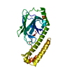 5e85C  5e86C 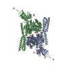 4jneS C: citing same article ( S: Starting model for refinement |
|---|---|
| Similar structure data |


-Links
 PDBj
PDBj


- Assembly
| Deposited unit | 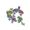
| ||||||||
|---|---|---|---|---|---|---|---|---|---|
| 1 | 
| ||||||||
| 2 | 
| ||||||||
| 3 | 
| ||||||||
| 4 | 
| ||||||||
| 5 | 
| ||||||||
| 6 | 
| ||||||||
| Unit cell |
|
-Components
-Protein , 1 types, 6 molecules ABCDEF
| #1: Protein | Mass: 67207.953 Da / Num. of mol.: 6 / Mutation: T229A, loop34 Source method: isolated from a genetically manipulated source Source: (gene. exp.) Homo sapiens (human) / Gene: HSPA5, GRP78 / Production host:  |
|---|
-Non-polymers , 5 types, 74 molecules 


| #2: Chemical | ChemComp-ATP /  Mass: 507.181 Da / Num. of mol.: 6 / Source method: obtained synthetically / Formula: C10H16N5O13P3 / Comment: ATP, energy-carrying molecule*YM Mass: 507.181 Da / Num. of mol.: 6 / Source method: obtained synthetically / Formula: C10H16N5O13P3 / Comment: ATP, energy-carrying molecule*YM#3: Chemical | ChemComp-ZN /  Mass: 65.409 Da / Num. of mol.: 18 / Source method: obtained synthetically / Formula: Zn Mass: 65.409 Da / Num. of mol.: 18 / Source method: obtained synthetically / Formula: Zn#4: Chemical | ChemComp-MG /  Mass: 24.305 Da / Num. of mol.: 12 / Source method: obtained synthetically / Formula: Mg Mass: 24.305 Da / Num. of mol.: 12 / Source method: obtained synthetically / Formula: Mg#5: Chemical | ChemComp-SO4 /  Mass: 96.063 Da / Num. of mol.: 6 / Source method: obtained synthetically / Formula: SO4 Mass: 96.063 Da / Num. of mol.: 6 / Source method: obtained synthetically / Formula: SO4#6: Water | ChemComp-HOH / | Mass: 18.015 Da / Num. of mol.: 32 / Source method: isolated from a natural source / Formula: H2O |
|---|
-Experimental details
-Experiment
| Experiment | Method: X-RAY DIFFRACTION |
|---|
- Sample preparation
| Crystal | Density Matthews: 3.72 Å3/Da / Density % sol: 66.97 % / Description: hexagonal plates |
|---|---|
| Crystal grow | Temperature: 277 K / Method: vapor diffusion, hanging drop / pH: 4.5 Details: 8%-12% PEG 3000, 0.1 M acetate acid, pH 4.5, and 0.2 M zinc acetate PH range: 4.5 |
-Data collection
| Diffraction | Mean temperature: 100 K |
|---|---|
| Diffraction source | Source: SYNCHROTRON / Site: NSLS / Beamline: X4C / Wavelength: 0.979 Å |
| Detector | Type: MAR CCD 165 mm / Detector: CCD / Date: Feb 8, 2013 |
| Radiation | Monochromator: focusing monochromator crystal / Protocol: SINGLE WAVELENGTH / Monochromatic (M) / Laue (L): M / Scattering type: x-ray |
| Radiation wavelength | Wavelength: 0.979 Å / Relative weight: 1 |
| Reflection | Resolution: 2.99→40.76 Å / Num. all: 111753 / Num. obs: 111753 / % possible obs: 97.8 % / Redundancy: 7.3 % / Rmerge(I) obs: 0.118 / Net I/σ(I): 25.4 |
| Reflection shell | Resolution: 3→3.05 Å / Redundancy: 7.3 % / Rmerge(I) obs: 0.402 / Mean I/σ(I) obs: 4.1 / % possible all: 98.6 |
- Processing
| Software |
| ||||||||||||||||||||||||||||||||||||||||||||||||||||||||||||||||||||||||||||||||||||||||||||||||||||||||||||||||||||||||||||||||||||||||||||||||||||||||||||||||||||||||||||||||||||||
|---|---|---|---|---|---|---|---|---|---|---|---|---|---|---|---|---|---|---|---|---|---|---|---|---|---|---|---|---|---|---|---|---|---|---|---|---|---|---|---|---|---|---|---|---|---|---|---|---|---|---|---|---|---|---|---|---|---|---|---|---|---|---|---|---|---|---|---|---|---|---|---|---|---|---|---|---|---|---|---|---|---|---|---|---|---|---|---|---|---|---|---|---|---|---|---|---|---|---|---|---|---|---|---|---|---|---|---|---|---|---|---|---|---|---|---|---|---|---|---|---|---|---|---|---|---|---|---|---|---|---|---|---|---|---|---|---|---|---|---|---|---|---|---|---|---|---|---|---|---|---|---|---|---|---|---|---|---|---|---|---|---|---|---|---|---|---|---|---|---|---|---|---|---|---|---|---|---|---|---|---|---|---|---|
| Refinement | Method to determine structure: MOLECULAR REPLACEMENT Starting model: 4JNE Resolution: 2.99→40.76 Å / Cor.coef. Fo:Fc: 0.89 / Cor.coef. Fo:Fc free: 0.855 / SU B: 19.146 / SU ML: 0.357 / Cross valid method: THROUGHOUT / ESU R Free: 0.444 / Stereochemistry target values: MAXIMUM LIKELIHOOD / Details: HYDROGENS HAVE BEEN ADDED IN THE RIDING POSITIONS
| ||||||||||||||||||||||||||||||||||||||||||||||||||||||||||||||||||||||||||||||||||||||||||||||||||||||||||||||||||||||||||||||||||||||||||||||||||||||||||||||||||||||||||||||||||||||
| Solvent computation | Ion probe radii: 0.8 Å / Shrinkage radii: 0.8 Å / VDW probe radii: 1.2 Å / Solvent model: MASK | ||||||||||||||||||||||||||||||||||||||||||||||||||||||||||||||||||||||||||||||||||||||||||||||||||||||||||||||||||||||||||||||||||||||||||||||||||||||||||||||||||||||||||||||||||||||
| Displacement parameters | Biso mean: 76.682 Å2
| ||||||||||||||||||||||||||||||||||||||||||||||||||||||||||||||||||||||||||||||||||||||||||||||||||||||||||||||||||||||||||||||||||||||||||||||||||||||||||||||||||||||||||||||||||||||
| Refinement step | Cycle: 1 / Resolution: 2.99→40.76 Å
| ||||||||||||||||||||||||||||||||||||||||||||||||||||||||||||||||||||||||||||||||||||||||||||||||||||||||||||||||||||||||||||||||||||||||||||||||||||||||||||||||||||||||||||||||||||||
| Refine LS restraints |
|