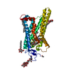
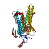
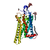
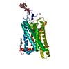
[English] 日本語
Yorodumi
- PDB-5dys: Crystal Structure of T94I rhodopsin mutant -

+
Open data


ID or keywords:

Loading...

-
Basic information

Entry
Database: PDB / ID: 5dys
TitleCrystal Structure of T94I rhodopsin mutant
ComponentsRhodopsin
KeywordsSIGNALING PROTEIN / Rhodopsin / G protein-coupled receptors / Congenital stationary night blindness / constitutive activity
Function / homology
Function and homology information


Opsins / VxPx cargo-targeting to cilium / rod photoreceptor outer segment / rod bipolar cell differentiation / sperm head plasma membrane / podosome assembly / absorption of visible light / opsin binding / The canonical retinoid cycle in rods (twilight vision) / : ...Opsins / VxPx cargo-targeting to cilium / rod photoreceptor outer segment / rod bipolar cell differentiation / sperm head plasma membrane / podosome assembly / absorption of visible light / opsin binding / The canonical retinoid cycle in rods (twilight vision) / : / G protein-coupled photoreceptor activity / photoreceptor inner segment membrane / rhodopsin mediated signaling pathway / 11-cis retinal binding / cellular response to light stimulus / G protein-coupled receptor complex / Inactivation, recovery and regulation of the phototransduction cascade / phototransduction, visible light / thermotaxis / Activation of the phototransduction cascade / detection of temperature stimulus involved in thermoception / outer membrane / arrestin family protein binding / photoreceptor cell maintenance / photoreceptor outer segment membrane / G alpha (i) signalling events / response to light stimulus / phototransduction / photoreceptor outer segment / G-protein alpha-subunit binding / sperm midpiece / visual perception / guanyl-nucleotide exchange factor activity / microtubule cytoskeleton organization / photoreceptor disc membrane / cell-cell junction / gene expression / G protein-coupled receptor signaling pathway / Golgi membrane / zinc ion binding / membrane / identical protein binding / plasma membrane
Similarity search - Function
Rhodopsin, N-terminal / Amino terminal of the G-protein receptor rhodopsin / Rhodopsin / Opsin / Visual pigments (opsins) retinal binding site / Visual pigments (opsins) retinal binding site. / Rhopdopsin 7-helix transmembrane proteins / Rhodopsin 7-helix transmembrane proteins / Serpentine type 7TM GPCR chemoreceptor Srsx / G-protein coupled receptors family 1 signature. ...Rhodopsin, N-terminal / Amino terminal of the G-protein receptor rhodopsin / Rhodopsin / Opsin / Visual pigments (opsins) retinal binding site / Visual pigments (opsins) retinal binding site. / Rhopdopsin 7-helix transmembrane proteins / Rhodopsin 7-helix transmembrane proteins / Serpentine type 7TM GPCR chemoreceptor Srsx / G-protein coupled receptors family 1 signature. / G protein-coupled receptor, rhodopsin-like / GPCR, rhodopsin-like, 7TM / G-protein coupled receptors family 1 profile. / 7 transmembrane receptor (rhodopsin family) / Up-down Bundle / Mainly Alpha
Similarity search - Domain/homology
ACETATE ION / PALMITIC ACID / RETINAL / Rhodopsin
Similarity search - Component
Biological speciesBos taurus (cattle)
MethodX-RAY DIFFRACTION / SYNCHROTRON / MOLECULAR REPLACEMENT / Resolution: 2.3 Å
AuthorsSinghal, A. / Guo, Y. / Matkovic, M. / Schertler, G. / Deupi, X. / Yan, E. / Standfuss, J.
Funding support Switzerland, 1items
OrganizationGrant numberCountry
Switzerland
CitationJournal: Embo Rep. / Year: 2016
Title: Structural role of the T94I rhodopsin mutation in congenital stationary night blindness.
Authors: Singhal, A. / Guo, Y. / Matkovic, M. / Schertler, G. / Deupi, X. / Yan, E.C. / Standfuss, J.
History
DepositionSep 25, 2015Deposition site: RCSB / Processing site: PDBE
Revision 1.0Aug 10, 2016Provider: repository / Type: Initial release
Revision 1.1Oct 19, 2016Group: Database references
Revision 2.0Jul 29, 2020Group: Atomic model / Data collection ...Atomic model / Data collection / Derived calculations / Structure summary
Category: atom_site / chem_comp ...atom_site / chem_comp / entity / pdbx_branch_scheme / pdbx_chem_comp_identifier / pdbx_entity_branch / pdbx_entity_branch_descriptor / pdbx_entity_branch_link / pdbx_entity_branch_list / pdbx_entity_nonpoly / pdbx_nonpoly_scheme / pdbx_struct_assembly_gen / struct_asym / struct_conn / struct_site / struct_site_gen
Item: _atom_site.B_iso_or_equiv / _atom_site.Cartn_x ..._atom_site.B_iso_or_equiv / _atom_site.Cartn_x / _atom_site.Cartn_y / _atom_site.Cartn_z / _atom_site.auth_asym_id / _atom_site.auth_atom_id / _atom_site.auth_comp_id / _atom_site.auth_seq_id / _atom_site.label_asym_id / _atom_site.label_atom_id / _atom_site.label_comp_id / _atom_site.label_entity_id / _atom_site.type_symbol / _chem_comp.mon_nstd_flag / _chem_comp.name / _chem_comp.type / _pdbx_struct_assembly_gen.asym_id_list / _struct_conn.pdbx_role / _struct_conn.ptnr1_auth_asym_id / _struct_conn.ptnr1_auth_seq_id / _struct_conn.ptnr1_label_asym_id / _struct_conn.ptnr2_auth_asym_id / _struct_conn.ptnr2_auth_seq_id / _struct_conn.ptnr2_label_asym_id
Description: Carbohydrate remediation / Provider: repository / Type: Remediation
Revision 2.1Jan 10, 2024Group: Data collection / Database references ...Data collection / Database references / Derived calculations / Refinement description / Structure summary
Category: chem_comp / chem_comp_atom ...chem_comp / chem_comp_atom / chem_comp_bond / database_2 / pdbx_initial_refinement_model / struct_conn
Item: _chem_comp.pdbx_synonyms / _database_2.pdbx_DOI ..._chem_comp.pdbx_synonyms / _database_2.pdbx_DOI / _database_2.pdbx_database_accession / _struct_conn.pdbx_leaving_atom_flag

-
Structure visualization

Structure viewerMolecule:
MolmilJmol/JSmol

Downloads & links

-
Assembly

Deposited unit
A: Rhodopsin
hetero molecules


Theoretical massNumber of molelcules
Total (without water)41,55410
Polymers39,0471
Non-polymers2,5079
Water52229
1


  • Idetical with deposited unit
  • defined by author&software
TypeNameSymmetry operationNumber
identity operation1_555x,y,z1
Buried area3760 Å2
ΔGint9 kcal/mol
Surface area17310 Å2
MethodPISA
Unit cell
Length a, b, c (Å)242.301, 242.301, 112.055
Angle α, β, γ (deg.)90.00, 90.00, 120.00
Int Tables number155
Space group name H-MH32

-
Components

-
Protein , 1 types, 1 molecules A

#1: Protein Rhodopsin /


Mass: 39046.641 Da / Num. of mol.: 1 / Mutation: N2C, D282C, T94I
Source method: isolated from a genetically manipulated source
Source: (gene. exp.) Bos taurus (cattle) / Gene: RHO / Plasmid: pCMV-tet O / Cell line (production host): HEK293S GNTI- / Production host: 
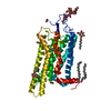
Homo sapiens (human) / References: UniProt: P02699

-
Sugars , 2 types, 3 molecules

#2: Polysaccharide alpha-D-mannopyranose-(1-3)-[beta-D-mannopyranose-(1-6)]beta-D-mannopyranose-(1-4)-2-acetamido-2- ...alpha-D-mannopyranose-(1-3)-[beta-D-mannopyranose-(1-6)]beta-D-mannopyranose-(1-4)-2-acetamido-2-deoxy-beta-D-glucopyranose-(1-4)-2-acetamido-2-deoxy-beta-D-glucopyranose


Type: oligosaccharide / Mass: 910.823 Da / Num. of mol.: 1
Source method: isolated from a genetically manipulated source
DescriptorTypeProgram
DManpa1-3[DManpb1-6]DManpb1-4DGlcpNAcb1-4DGlcpNAcb1-Glycam Condensed SequenceGMML 1.0
WURCS=2.0/3,5,4/[a2122h-1b_1-5_2*NCC/3=O][a1122h-1b_1-5][a1122h-1a_1-5]/1-1-2-3-2/a4-b1_b4-c1_c3-d1_c6-e1WURCSPDB2Glycan 1.1.0
[]{[(4+1)][b-D-GlcpNAc]{[(4+1)][b-D-GlcpNAc]{[(4+1)][b-D-Manp]{[(3+1)][a-D-Manp]{}[(6+1)][b-D-Manp]{}}}}}LINUCSPDB-CARE
#5: Sugar ChemComp-BOG / octyl beta-D-glucopyranoside / Beta-Octylglucoside / octyl beta-D-glucoside / octyl D-glucoside / octyl glucoside / Octyl glucoside


Type: D-saccharide / Mass: 292.369 Da / Num. of mol.: 2
Source method: isolated from a genetically manipulated source
Formula: C14H28O6 / Comment: detergent*YM
IdentifierTypeProgram
b-octylglucosideIUPAC CARBOHYDRATE SYMBOLPDB-CARE 1.0

-
Non-polymers , 5 types, 35 molecules

#3: Chemical ChemComp-RET / RETINAL / Retinal


Mass: 284.436 Da / Num. of mol.: 1 / Mutation: N2C, D282C, T94I
Source method: isolated from a genetically manipulated source
Formula: C20H28O
#4: Chemical ChemComp-ACT / ACETATE ION / Acetate


Mass: 59.044 Da / Num. of mol.: 2 / Source method: obtained synthetically / Formula: C2H3O2
#6: Chemical ChemComp-PLM / PALMITIC ACID / Palmitic acid


Mass: 256.424 Da / Num. of mol.: 2 / Source method: obtained synthetically / Formula: C16H32O2
#7: Chemical ChemComp-SO4 / SULFATE ION / Sulfate


Mass: 96.063 Da / Num. of mol.: 1 / Source method: obtained synthetically / Formula: SO4
#8: Water ChemComp-HOH / water / Water


Mass: 18.015 Da / Num. of mol.: 29 / Source method: isolated from a natural source / Formula: H2O

-
Experimental details

-
Experiment

ExperimentMethod: X-RAY DIFFRACTION

-
Sample preparation

Crystal growTemperature: 277 K / Method: vapor diffusion, hanging drop / Details: Ammonium Sulfate, Sodium acetate / PH range: 4-6

-
Data collection

DiffractionMean temperature: 100 K
Diffraction sourceSource: SYNCHROTRON / Site: SLS / Beamline: X06DA / Wavelength: 1 Å
DetectorType: PSI PILATUS 6M / Detector: PIXEL / Date: Oct 17, 2012
RadiationProtocol: SINGLE WAVELENGTH / Monochromatic (M) / Laue (L): M / Scattering type: x-ray
Radiation wavelengthWavelength: 1 Å / Relative weight: 1
ReflectionResolution: 2.3→49.4 Å / Num. obs: 55751 / % possible obs: 99.7 % / Redundancy: 18.9 % / Rmerge(I) obs: 0.094 / Net I/σ(I): 20.11
Reflection shellHighest resolution: 2.3 Å / Rmerge(I) obs: 2.243

-
Processing

Software
NameVersionClassification
REFMAC5.8.0131refinement
XDSdata reduction
XSCALEdata scaling
MOLREPphasing
RefinementMethod to determine structure: MOLECULAR REPLACEMENT
Starting model: 4A4M
Resolution: 2.3→49.4 Å / Cor.coef. Fo:Fc: 0.94 / Cor.coef. Fo:Fc free: 0.939 / SU B: 6.013 / SU ML: 0.128 / Cross valid method: THROUGHOUT / ESU R: 0.134 / ESU R Free: 0.125 / Stereochemistry target values: MAXIMUM LIKELIHOOD / Details: HYDROGENS HAVE BEEN ADDED IN THE RIDING POSITIONS
RfactorNum. reflection% reflectionSelection details
Rfree0.231 2776 5 %RANDOM
Rwork0.21936 ---
obs0.21995 52726 99.77 %-
Solvent computationIon probe radii: 0.8 Å / Shrinkage radii: 0.8 Å / VDW probe radii: 1.2 Å / Solvent model: MASK
Displacement parametersBiso mean: 66.093 Å2
Baniso -1Baniso -2Baniso -3
1--2.7 Å2-1.35 Å20 Å2
2---2.7 Å20 Å2
3---8.75 Å2
Refinement stepCycle: 1 / Resolution: 2.3→49.4 Å
ProteinNucleic acidLigandSolventTotal
Num. atoms2592 0 168 29 2789
Refine LS restraints
Refine-IDTypeDev idealDev ideal targetNumber
X-RAY DIFFRACTIONr_bond_refined_d0.0140.022878
X-RAY DIFFRACTIONr_bond_other_d0.0020.022724
X-RAY DIFFRACTIONr_angle_refined_deg1.7972.0013918
X-RAY DIFFRACTIONr_angle_other_deg1.04736280
X-RAY DIFFRACTIONr_dihedral_angle_1_deg6.1675331
X-RAY DIFFRACTIONr_dihedral_angle_2_deg32.91822.991112
X-RAY DIFFRACTIONr_dihedral_angle_3_deg16.05915415
X-RAY DIFFRACTIONr_dihedral_angle_4_deg20.137157
X-RAY DIFFRACTIONr_chiral_restr0.0940.2444
X-RAY DIFFRACTIONr_gen_planes_refined0.010.0213096
X-RAY DIFFRACTIONr_gen_planes_other0.0020.02685
X-RAY DIFFRACTIONr_nbd_refined
X-RAY DIFFRACTIONr_nbd_other
X-RAY DIFFRACTIONr_nbtor_refined
X-RAY DIFFRACTIONr_nbtor_other
X-RAY DIFFRACTIONr_xyhbond_nbd_refined
X-RAY DIFFRACTIONr_xyhbond_nbd_other
X-RAY DIFFRACTIONr_metal_ion_refined
X-RAY DIFFRACTIONr_metal_ion_other
X-RAY DIFFRACTIONr_symmetry_vdw_refined
X-RAY DIFFRACTIONr_symmetry_vdw_other
X-RAY DIFFRACTIONr_symmetry_hbond_refined
X-RAY DIFFRACTIONr_symmetry_hbond_other
X-RAY DIFFRACTIONr_symmetry_metal_ion_refined
X-RAY DIFFRACTIONr_symmetry_metal_ion_other
X-RAY DIFFRACTIONr_mcbond_it4.6816.3031316
X-RAY DIFFRACTIONr_mcbond_other4.6576.2921313
X-RAY DIFFRACTIONr_mcangle_it6.6469.4331643
X-RAY DIFFRACTIONr_mcangle_other6.6449.4381644
X-RAY DIFFRACTIONr_scbond_it5.556.91562
X-RAY DIFFRACTIONr_scbond_other5.4746.8951558
X-RAY DIFFRACTIONr_scangle_it
X-RAY DIFFRACTIONr_scangle_other8.13310.1272268
X-RAY DIFFRACTIONr_long_range_B_refined9.39451.7183204
X-RAY DIFFRACTIONr_long_range_B_other9.39951.7233199
X-RAY DIFFRACTIONr_rigid_bond_restr
X-RAY DIFFRACTIONr_sphericity_free
X-RAY DIFFRACTIONr_sphericity_bonded
LS refinement shellResolution: 2.301→2.36 Å / Total num. of bins used: 20
RfactorNum. reflection% reflection
Rfree0.429 198 -
Rwork0.445 3758 -
obs--97.1 %

+
About Yorodumi

-
News

-
Feb 9, 2022. New format data for meta-information of EMDB entries

New format data for meta-information of EMDB entries

  • Version 3 of the EMDB header file is now the official format.
  • The previous official version 1.9 will be removed from the archive.

Related info.:EMDB header

External links:wwPDB to switch to version 3 of the EMDB data model

-
Aug 12, 2020. Covid-19 info

Covid-19 info

URL: https://pdbj.org/emnavi/covid19.php

New page: Covid-19 featured information page in EM Navigator.

Related info.:Covid-19 info / Mar 5, 2020. Novel coronavirus structure data

+
Mar 5, 2020. Novel coronavirus structure data

Novel coronavirus structure data

Related info.:Yorodumi Speices / Aug 12, 2020. Covid-19 info

External links:COVID-19 featured content - PDBj / Molecule of the Month (242):Coronavirus Proteases

+
Jan 31, 2019. EMDB accession codes are about to change! (news from PDBe EMDB page)

EMDB accession codes are about to change! (news from PDBe EMDB page)

  • The allocation of 4 digits for EMDB accession codes will soon come to an end. Whilst these codes will remain in use, new EMDB accession codes will include an additional digit and will expand incrementally as the available range of codes is exhausted. The current 4-digit format prefixed with “EMD-” (i.e. EMD-XXXX) will advance to a 5-digit format (i.e. EMD-XXXXX), and so on. It is currently estimated that the 4-digit codes will be depleted around Spring 2019, at which point the 5-digit format will come into force.
  • The EM Navigator/Yorodumi systems omit the EMD- prefix.

Related info.:Q: What is EMD? / ID/Accession-code notation in Yorodumi/EM Navigator

External links:EMDB Accession Codes are Changing Soon! / Contact to PDBj

+
Jul 12, 2017. Major update of PDB

Major update of PDB

  • wwPDB released updated PDB data conforming to the new PDBx/mmCIF dictionary.
  • This is a major update changing the version number from 4 to 5, and with Remediation, in which all the entries are updated.
  • In this update, many items about electron microscopy experimental information are reorganized (e.g. em_software).
  • Now, EM Navigator and Yorodumi are based on the updated data.

External links:wwPDB Remediation / Enriched Model Files Conforming to OneDep Data Standards Now Available in the PDB FTP Archive

-
Yorodumi

Thousand views of thousand structures

  • Yorodumi is a browser for structure data from EMDB, PDB, SASBDB, etc.
  • This page is also the successor to EM Navigator detail page, and also detail information page/front-end page for Omokage search.
  • The word "yorodu" (or yorozu) is an old Japanese word meaning "ten thousand". "mi" (miru) is to see.

Related info.:EMDB / PDB / SASBDB / Comparison of 3 databanks / Yorodumi Search / Aug 31, 2016. New EM Navigator & Yorodumi / Yorodumi Papers / Jmol/JSmol / Function and homology information / Changes in new EM Navigator and Yorodumi

Read more