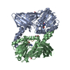
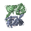
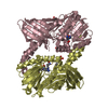
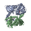
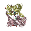
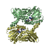
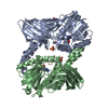
Entry Database : PDB / ID : 5dx0Title Crystal structure of CARM1, sinefungin, and H3 peptide (R17) H3 peptide Histone-arginine methyltransferase CARM1 Keywords / Function / homology Function Domain/homology Component
/ / / / / / / / / / / / / / / / / / / / / / / / / / / / / / / / / / / / / / / / / / / / / / / / / / / / / / / / / / / / / / / / / / / / / / / / / / / / / / / / / / / / / / / / / / / / / / / / / / / / / / / / / / / / / / / / / / / / / / / / / / / / / / / / / Biological species Homo sapiens (human)Method / / Resolution : 2.05 Å Authors Boriack-Sjodin, P.A. Journal : Acs Chem.Biol. / Year : 2016Title : Structural Insights into Ternary Complex Formation of Human CARM1 with Various Substrates.Authors : Boriack-Sjodin, P.A. / Jin, L. / Jacques, S.L. / Drew, A. / Sneeringer, C. / Scott, M.P. / Moyer, M.P. / Ribich, S. / Moradei, O. / Copeland, R.A. History Deposition Sep 23, 2015 Deposition site / Processing site Revision 1.0 Nov 25, 2015 Provider / Type Revision 1.1 Mar 30, 2016 Group Revision 1.2 Oct 30, 2024 Group Data collection / Database references ... Data collection / Database references / Derived calculations / Structure summary Category chem_comp_atom / chem_comp_bond ... chem_comp_atom / chem_comp_bond / database_2 / pdbx_entry_details / pdbx_modification_feature / pdbx_struct_oper_list Item / _database_2.pdbx_database_accession / _pdbx_struct_oper_list.symmetry_operation
Show all Show less
 Movie
Movie Controller


 Open data
 Basic information
 Components
 Keywords
 Function and homology information
 Homo sapiens (human)
 X-RAY DIFFRACTION /
  Authors
 Citation
 Structure visualization
 Downloads & links
 Other downloads


 PDBj
PDBj


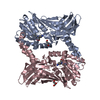
 Assembly
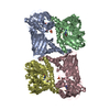


 Spodoptera frugiperda (fall armyworm)


 Mass: 381.387 Da / Num. of mol.: 4 / Source method: obtained synthetically / Formula: C15H23N7O5
 Mass: 92.094 Da / Num. of mol.: 2 / Source method: obtained synthetically / Formula: C3H8O3
 Mass: 96.063 Da / Num. of mol.: 4 / Source method: obtained synthetically / Formula: SO4
 Sample preparation
 / Beamline: BL17U / Wavelength: 0.97915 Å
 Processing