Polo-like kinase mediated events / Phosphorylation of the APC/C / Phosphorylation of Emi1 / Mitotic Metaphase/Anaphase Transition / Activation of NIMA Kinases NEK9, NEK6, NEK7 / Golgi Cisternae Pericentriolar Stack Reorganization / Mitotic Telophase/Cytokinesis / Cyclin A/B1/B2 associated events during G2/M transition / Condensation of Prophase Chromosomes / polar body extrusion after meiotic divisions ...Polo-like kinase mediated events / Phosphorylation of the APC/C / Phosphorylation of Emi1 / Mitotic Metaphase/Anaphase Transition / Activation of NIMA Kinases NEK9, NEK6, NEK7 / Golgi Cisternae Pericentriolar Stack Reorganization / Mitotic Telophase/Cytokinesis / Cyclin A/B1/B2 associated events during G2/M transition / Condensation of Prophase Chromosomes / polar body extrusion after meiotic divisions / centrosome separation / protein localization to nuclear envelope / APC/C:Cdh1 mediated degradation of Cdc20 and other APC/C:Cdh1 targeted proteins in late mitosis/early G1 / Amplification of signal from unattached kinetochores via a MAD2 inhibitory signal / regulation of protein localization to cell cortex / nuclear membrane disassembly / synaptonemal complex disassembly / Mitotic Prometaphase / EML4 and NUDC in mitotic spindle formation / Resolution of Sister Chromatid Cohesion / The role of GTSE1 in G2/M progression after G2 checkpoint / polo kinase / RHO GTPases Activate Formins / homologous chromosome segregation / Loss of Nlp from mitotic centrosomes / Recruitment of mitotic centrosome proteins and complexes / Loss of proteins required for interphase microtubule organization from the centrosome / Anchoring of the basal body to the plasma membrane / Separation of Sister Chromatids / Recruitment of NuMA to mitotic centrosomes / AURKA Activation by TPX2 / female meiosis chromosome segregation / anaphase-promoting complex binding / synaptonemal complex / Regulation of PLK1 Activity at G2/M Transition / mitotic cleavage furrow formation / outer kinetochore / condensed chromosome, centromeric region / microtubule bundle formation / double-strand break repair via alternative nonhomologous end joining / regulation of mitotic spindle assembly / protein localization to site of double-strand break / positive regulation of mitotic metaphase/anaphase transition / protein localization to centrosome / centrosome cycle / regulation of mitotic metaphase/anaphase transition / mitotic spindle pole / mitotic spindle assembly checkpoint signaling / spindle midzone / establishment of mitotic spindle orientation / mitotic G2 DNA damage checkpoint signaling / positive regulation of proteolysis / mitotic cytokinesis / mitotic sister chromatid segregation / chromosome, centromeric region / negative regulation of double-strand break repair via homologous recombination / protein localization to chromatin / regulation of mitotic cell cycle / mitotic spindle organization / regulation of cytokinesis / spindle microtubule / establishment of protein localization / centriole / kinetochore / double-strand break repair via homologous recombination / protein destabilization / G2/M transition of mitotic cell cycle / spindle / centriolar satellite / spindle pole / double-strand break repair / mitotic cell cycle / positive regulation of proteasomal ubiquitin-dependent protein catabolic process / microtubule cytoskeleton / midbody / microtubule binding / protein phosphorylation / protein kinase activity / protein ubiquitination / protein serine kinase activity / protein serine/threonine kinase activity / centrosome / protein kinase binding / negative regulation of apoptotic process / chromatin / magnesium ion binding / negative regulation of transcription by RNA polymerase II / ATP binding / identical protein binding / nucleus / cytoplasm Similarity search - Function
POLO box domain / Polo-like kinase 1, catalytic domain / Second polo-box domain / First polo-box domain / POLO box domain superfamily / POLO box duplicated region / Arylsulfatase, C-terminal domain / POLO box domain / POLO box domain profile. / Serine/threonine-protein kinase, active site ...POLO box domain / Polo-like kinase 1, catalytic domain / Second polo-box domain / First polo-box domain / POLO box domain superfamily / POLO box duplicated region / Arylsulfatase, C-terminal domain / POLO box domain / POLO box domain profile. / Serine/threonine-protein kinase, active site / Serine/Threonine protein kinases active-site signature. / Protein kinase domain / Serine/Threonine protein kinases, catalytic domain / Protein kinase, ATP binding site / Protein kinases ATP-binding region signature. / Protein kinase domain profile. / Protein kinase domain / Protein kinase-like domain superfamily / 2-Layer Sandwich / Alpha Beta Similarity search - Domain/homology
Monochromator: DCM Si (111) Crystal / Protocol: SINGLE WAVELENGTH / Monochromatic (M) / Laue (L): M / Scattering type: x-ray
Radiation wavelength
Wavelength: 1 Å / Relative weight: 1
Reflection
Resolution: 2.3→44.7 Å / Num. obs: 9156 / % possible obs: 90.4 % / Redundancy: 3.6 % / Net I/σ(I): 23.4
-
Processing
Software
Name
Version
Classification
REFMAC
5.8.0107
refinement
HKL-2000
datareduction
HKL-2000
datascaling
PHASER
phasing
Refinement
Resolution: 2.3→44.7 Å / Cor.coef. Fo:Fc: 0.94 / Cor.coef. Fo:Fc free: 0.878 / SU B: 8.572 / SU ML: 0.205 / Cross valid method: THROUGHOUT / ESU R: 0.444 / ESU R Free: 0.296 / Stereochemistry target values: MAXIMUM LIKELIHOOD / Details: HYDROGENS HAVE BEEN ADDED IN THE RIDING POSITIONS
Rfactor
Num. reflection
% reflection
Selection details
Rfree
0.27693
466
4.8 %
RANDOM
Rwork
0.19603
-
-
-
obs
0.19983
9156
90.48 %
-
Solvent computation
Ion probe radii: 0.8 Å / Shrinkage radii: 0.8 Å / VDW probe radii: 1.2 Å / Solvent model: MASK
 Movie
Movie Controller


 Open data
 Basic information
 Components
 Keywords
 Function and homology information
 X-RAY DIFFRACTION /
  Authors
 Citation
 Structure visualization
 Downloads & links
 Other downloads
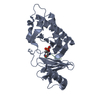


 PDBj
PDBj


 Assembly
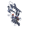


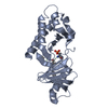
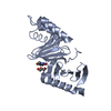
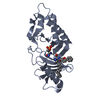
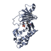
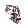
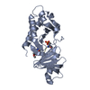
 Type: Polypeptide / Class: Enzyme inhibitor / Mass: 893.738 Da / Num. of mol.: 1 / Source method: obtained synthetically / Source: (synth.) synthetic construct (others) / References: peptide inhibitor
 Mass: 18.015 Da / Num. of mol.: 11 / Source method: isolated from a natural source / Formula: H2O
 Sample preparation
 / Beamline: 7A (6B, 6C1) / Wavelength: 1 Å
 Processing