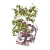
[English] 日本語
Yorodumi
- PDB-5dgq: Crystal structure of GH9 exo-beta-D-glucosaminidase PBPRA0520 -

+
Open data


ID or keywords:

Loading...

-
Basic information

Entry
Database: PDB / ID: 5dgq
TitleCrystal structure of GH9 exo-beta-D-glucosaminidase PBPRA0520
ComponentsPutative endoglucanase-related protein
KeywordsHYDROLASE / Glycoside Hydrolase family 9 / alpha-alpha-6 barrel / exo-D-beta-glucosaminidase
Function / homology
Function and homology information


cellulase activity / polysaccharide catabolic process
Similarity search - Function
Cellulase, Ig-like domain / Glycoside hydrolase family 9 / Glycosyl hydrolase family 9 / Glycosyltransferase - #10 / Six-hairpin glycosidase-like superfamily / Six-hairpin glycosidase superfamily / Glycosyltransferase / Alpha/alpha barrel / Immunoglobulin E-set / Immunoglobulins ...Cellulase, Ig-like domain / Glycoside hydrolase family 9 / Glycosyl hydrolase family 9 / Glycosyltransferase - #10 / Six-hairpin glycosidase-like superfamily / Six-hairpin glycosidase superfamily / Glycosyltransferase / Alpha/alpha barrel / Immunoglobulin E-set / Immunoglobulins / Immunoglobulin-like fold / Immunoglobulin-like / Sandwich / Mainly Beta / Mainly Alpha
Similarity search - Domain/homology
Endoglucanase-related protein
Similarity search - Component
Biological speciesPhotobacterium profundum (bacteria)
MethodX-RAY DIFFRACTION / SYNCHROTRON / MOLECULAR REPLACEMENT / Resolution: 1.9 Å
AuthorsSuzuki, K. / Honda, Y. / Fushinobu, S.
Funding support Japan, 1items
OrganizationGrant numberCountry
Ministry of Education, Culture, Sports, Science and Technology (Japan)22780095 Japan
Citation
Journal: Biochem.J. / Year: 2016
Title: The crystal structure of an inverting glycoside hydrolase family 9 exo-beta-D-glucosaminidase and the design of glycosynthase.
Authors: Honda, Y. / Arai, S. / Suzuki, K. / Kitaoka, M. / Fushinobu, S.
#1: Journal: Glycobiology / Year: 2011
Title: Elucidation of exo-beta-D-glucosaminidase activity of a family 9 glycoside hydrolase (PBPRA0520) from Photobacterium profundum SS9.
Authors: Honda, Y. / Shimaya, N. / Ishisaki, K. / Ebihara, M. / Taniguchi, H.
History
DepositionAug 28, 2015Deposition site: RCSB / Processing site: PDBJ
Revision 1.0Dec 9, 2015Provider: repository / Type: Initial release
Revision 1.1Apr 20, 2016Group: Database references
Revision 1.2Feb 19, 2020Group: Author supporting evidence / Data collection ...Author supporting evidence / Data collection / Database references / Derived calculations
Category: citation / diffrn_source ...citation / diffrn_source / pdbx_audit_support / pdbx_struct_oper_list
Item: _citation.journal_id_CSD / _diffrn_source.pdbx_synchrotron_site ..._citation.journal_id_CSD / _diffrn_source.pdbx_synchrotron_site / _pdbx_audit_support.funding_organization / _pdbx_struct_oper_list.symmetry_operation
Revision 1.3Nov 8, 2023Group: Data collection / Database references ...Data collection / Database references / Derived calculations / Refinement description
Category: chem_comp_atom / chem_comp_bond ...chem_comp_atom / chem_comp_bond / database_2 / pdbx_initial_refinement_model / pdbx_struct_conn_angle / struct_conn
Item: _database_2.pdbx_DOI / _database_2.pdbx_database_accession ..._database_2.pdbx_DOI / _database_2.pdbx_database_accession / _pdbx_struct_conn_angle.ptnr1_auth_seq_id / _pdbx_struct_conn_angle.ptnr3_auth_seq_id / _pdbx_struct_conn_angle.value / _struct_conn.pdbx_dist_value / _struct_conn.ptnr1_auth_asym_id / _struct_conn.ptnr1_auth_comp_id / _struct_conn.ptnr1_auth_seq_id / _struct_conn.ptnr1_label_asym_id / _struct_conn.ptnr1_label_atom_id / _struct_conn.ptnr1_label_comp_id / _struct_conn.ptnr1_label_seq_id / _struct_conn.ptnr2_auth_asym_id / _struct_conn.ptnr2_auth_comp_id / _struct_conn.ptnr2_auth_seq_id / _struct_conn.ptnr2_label_asym_id / _struct_conn.ptnr2_label_atom_id / _struct_conn.ptnr2_label_comp_id

-
Structure visualization

Structure viewerMolecule:
MolmilJmol/JSmol

Downloads & links

-
Assembly

Deposited unit
A: Putative endoglucanase-related protein
B: Putative endoglucanase-related protein
hetero molecules


Theoretical massNumber of molelcules
Total (without water)133,3194
Polymers133,2732
Non-polymers462
Water14,772820
1


  • Idetical with deposited unit
  • defined by author&software
TypeNameSymmetry operationNumber
identity operation1_555x,y,z1
Buried area3270 Å2
ΔGint-48 kcal/mol
Surface area38330 Å2
MethodPISA
Unit cell
Length a, b, c (Å)61.928, 102.318, 90.280
Angle α, β, γ (deg.)90.00, 97.72, 90.00
Int Tables number4
Space group name H-MP1211

-
Components

#1: Protein Putative endoglucanase-related protein / exo-beta-D-glucosaminidase


Mass: 66636.633 Da / Num. of mol.: 2
Source method: isolated from a genetically manipulated source
Source: (gene. exp.) Photobacterium profundum (bacteria) / Gene: VV2739, PBPRA0520 / Plasmid: pET30 / Production host: Escherichia coli (E. coli) / Strain (production host): BL21(DE3) / References: UniProt: Q6LUT2
#2: Chemical ChemComp-NA / SODIUM ION


Mass: 22.990 Da / Num. of mol.: 2 / Source method: obtained synthetically / Formula: Na
#3: Water ChemComp-HOH / water


Mass: 18.015 Da / Num. of mol.: 820 / Source method: isolated from a natural source / Formula: H2O

-
Experimental details

-
Experiment

ExperimentMethod: X-RAY DIFFRACTION

-
Sample preparation

CrystalDensity Matthews: 2.16 Å3/Da / Density % sol: 43.2 %
Crystal growTemperature: 298 K / Method: vapor diffusion, sitting drop / pH: 7.5 / Details: 10% PEG 6000, 0.1M HEPES-NaOH (pH 7.5), 5% MPD

-
Data collection

DiffractionMean temperature: 100 K
Diffraction sourceSource: SYNCHROTRON / Site: Photon Factory / Beamline: BL-5A / Wavelength: 1 Å
DetectorType: ADSC QUANTUM 315r / Detector: CCD / Date: Dec 20, 2010
RadiationMonochromator: Numerical link type Si(111) double crystal monochromator
Protocol: SINGLE WAVELENGTH / Monochromatic (M) / Laue (L): M / Scattering type: x-ray
Radiation wavelengthWavelength: 1 Å / Relative weight: 1
ReflectionResolution: 1.9→50 Å / Num. obs: 87933 / % possible obs: 99.7 % / Redundancy: 3.7 % / Rmerge(I) obs: 0.1 / Net I/σ(I): 15.3
Reflection shellResolution: 1.9→1.97 Å / Redundancy: 3.8 % / Rmerge(I) obs: 0.289 / Mean I/σ(I) obs: 3.5 / % possible all: 100

-
Processing

Software
NameVersionClassification
REFMAC5.8.0107refinement
HKL-2000data reduction
HKL-2000data scaling
MOLREPphasing
RefinementMethod to determine structure: MOLECULAR REPLACEMENT
Starting model: 3H7L
Resolution: 1.9→31.87 Å / Cor.coef. Fo:Fc: 0.961 / Cor.coef. Fo:Fc free: 0.937 / SU B: 3.052 / SU ML: 0.091 / Cross valid method: THROUGHOUT / ESU R: 0.146 / ESU R Free: 0.138 / Stereochemistry target values: MAXIMUM LIKELIHOOD / Details: HYDROGENS HAVE BEEN USED IF PRESENT IN THE INPUT
RfactorNum. reflection% reflectionSelection details
Rfree0.20625 4408 5 %RANDOM
Rwork0.15728 ---
obs0.15972 83390 98.99 %-
Solvent computationIon probe radii: 0.8 Å / Shrinkage radii: 0.8 Å / VDW probe radii: 1.2 Å / Solvent model: MASK
Displacement parametersBiso mean: 20.004 Å2
Baniso -1Baniso -2Baniso -3
1--0.09 Å20 Å2-0.11 Å2
2--0 Å20 Å2
3---0.11 Å2
Refinement stepCycle: 1 / Resolution: 1.9→31.87 Å
ProteinNucleic acidLigandSolventTotal
Num. atoms9183 0 2 820 10005
Refine LS restraints
Refine-IDTypeDev idealDev ideal targetNumber
X-RAY DIFFRACTIONr_bond_refined_d0.0230.0199421
X-RAY DIFFRACTIONr_bond_other_d
X-RAY DIFFRACTIONr_angle_refined_deg1.9781.92212776
X-RAY DIFFRACTIONr_angle_other_deg
X-RAY DIFFRACTIONr_dihedral_angle_1_deg6.77751148
X-RAY DIFFRACTIONr_dihedral_angle_2_deg35.25124.677496
X-RAY DIFFRACTIONr_dihedral_angle_3_deg13.827151521
X-RAY DIFFRACTIONr_dihedral_angle_4_deg17.0031544
X-RAY DIFFRACTIONr_chiral_restr0.1540.21322
X-RAY DIFFRACTIONr_gen_planes_refined0.0120.0217396
X-RAY DIFFRACTIONr_gen_planes_other
X-RAY DIFFRACTIONr_nbd_refined
X-RAY DIFFRACTIONr_nbd_other
X-RAY DIFFRACTIONr_nbtor_refined
X-RAY DIFFRACTIONr_nbtor_other
X-RAY DIFFRACTIONr_xyhbond_nbd_refined
X-RAY DIFFRACTIONr_xyhbond_nbd_other
X-RAY DIFFRACTIONr_metal_ion_refined
X-RAY DIFFRACTIONr_metal_ion_other
X-RAY DIFFRACTIONr_symmetry_vdw_refined
X-RAY DIFFRACTIONr_symmetry_vdw_other
X-RAY DIFFRACTIONr_symmetry_hbond_refined
X-RAY DIFFRACTIONr_symmetry_hbond_other
X-RAY DIFFRACTIONr_symmetry_metal_ion_refined
X-RAY DIFFRACTIONr_symmetry_metal_ion_other
X-RAY DIFFRACTIONr_mcbond_it1.781.7944601
X-RAY DIFFRACTIONr_mcbond_other
X-RAY DIFFRACTIONr_mcangle_it2.4152.6775746
X-RAY DIFFRACTIONr_mcangle_other
X-RAY DIFFRACTIONr_scbond_it2.9622.0214820
X-RAY DIFFRACTIONr_scbond_other
X-RAY DIFFRACTIONr_scangle_it
X-RAY DIFFRACTIONr_scangle_other
X-RAY DIFFRACTIONr_long_range_B_refined5.52815.73716032
X-RAY DIFFRACTIONr_long_range_B_other
X-RAY DIFFRACTIONr_rigid_bond_restr
X-RAY DIFFRACTIONr_sphericity_free
X-RAY DIFFRACTIONr_sphericity_bonded
LS refinement shellResolution: 1.893→1.942 Å / Total num. of bins used: 20
RfactorNum. reflection% reflection
Rfree0.254 308 -
Rwork0.19 5652 -
obs--91.79 %

+
About Yorodumi

-
News

-
Feb 9, 2022. New format data for meta-information of EMDB entries

New format data for meta-information of EMDB entries

  • Version 3 of the EMDB header file is now the official format.
  • The previous official version 1.9 will be removed from the archive.

Related info.:EMDB header

External links:wwPDB to switch to version 3 of the EMDB data model

-
Aug 12, 2020. Covid-19 info

Covid-19 info

URL: https://pdbjlvh1.pdbj.org/emnavi/covid19.php

New page: Covid-19 featured information page in EM Navigator.

Related info.:Covid-19 info / Mar 5, 2020. Novel coronavirus structure data

+
Mar 5, 2020. Novel coronavirus structure data

Novel coronavirus structure data

Related info.:Yorodumi Speices / Aug 12, 2020. Covid-19 info

External links:COVID-19 featured content - PDBj / Molecule of the Month (242):Coronavirus Proteases

+
Jan 31, 2019. EMDB accession codes are about to change! (news from PDBe EMDB page)

EMDB accession codes are about to change! (news from PDBe EMDB page)

  • The allocation of 4 digits for EMDB accession codes will soon come to an end. Whilst these codes will remain in use, new EMDB accession codes will include an additional digit and will expand incrementally as the available range of codes is exhausted. The current 4-digit format prefixed with “EMD-” (i.e. EMD-XXXX) will advance to a 5-digit format (i.e. EMD-XXXXX), and so on. It is currently estimated that the 4-digit codes will be depleted around Spring 2019, at which point the 5-digit format will come into force.
  • The EM Navigator/Yorodumi systems omit the EMD- prefix.

Related info.:Q: What is EMD? / ID/Accession-code notation in Yorodumi/EM Navigator

External links:EMDB Accession Codes are Changing Soon! / Contact to PDBj

+
Jul 12, 2017. Major update of PDB

Major update of PDB

  • wwPDB released updated PDB data conforming to the new PDBx/mmCIF dictionary.
  • This is a major update changing the version number from 4 to 5, and with Remediation, in which all the entries are updated.
  • In this update, many items about electron microscopy experimental information are reorganized (e.g. em_software).
  • Now, EM Navigator and Yorodumi are based on the updated data.

External links:wwPDB Remediation / Enriched Model Files Conforming to OneDep Data Standards Now Available in the PDB FTP Archive

-
Yorodumi

Thousand views of thousand structures

  • Yorodumi is a browser for structure data from EMDB, PDB, SASBDB, etc.
  • This page is also the successor to EM Navigator detail page, and also detail information page/front-end page for Omokage search.
  • The word "yorodu" (or yorozu) is an old Japanese word meaning "ten thousand". "mi" (miru) is to see.

Related info.:EMDB / PDB / SASBDB / Comparison of 3 databanks / Yorodumi Search / Aug 31, 2016. New EM Navigator & Yorodumi / Yorodumi Papers / Jmol/JSmol / Function and homology information / Changes in new EM Navigator and Yorodumi

Read more