 Movie
Movie Controller

[English] 日本語
 Yorodumi
- PDB-5chc: Crystal structure of the perchlorate reductase PcrAB - substrate ... -
+ Open data

- Basic information
| Entry | Database: PDB / ID: 5chc | ||||||
|---|---|---|---|---|---|---|---|
| Title | Crystal structure of the perchlorate reductase PcrAB - substrate analog SeO3 bound - from Azospira suillum PS | ||||||
 Components Components | (DMSO reductase family type II enzyme, ...) x 2 | ||||||
 Keywords Keywords | OXIDOREDUCTASE / electron-shuttling protein | ||||||
| Function / homology |  Function and homology information Function and homology informationoxidoreductase complex / cell envelope / molybdopterin cofactor binding / anaerobic respiration / cellular respiration / 3 iron, 4 sulfur cluster binding / 4 iron, 4 sulfur cluster binding / electron transfer activity / periplasmic space / oxidoreductase activity ...oxidoreductase complex / cell envelope / molybdopterin cofactor binding / anaerobic respiration / cellular respiration / 3 iron, 4 sulfur cluster binding / 4 iron, 4 sulfur cluster binding / electron transfer activity / periplasmic space / oxidoreductase activity / membrane / metal ion binding Similarity search - Function | ||||||
| Biological species |  Azospira oryzae (bacteria) Azospira oryzae (bacteria) | ||||||
| Method |  X-RAY DIFFRACTION / SYNCHROTRON / MOLECULAR REPLACEMENT / Resolution: 2.38 Å X-RAY DIFFRACTION / SYNCHROTRON / MOLECULAR REPLACEMENT / Resolution: 2.38 Å | ||||||
 Authors Authors | Tsai, C.-L. / Tainer, J.A. | ||||||
 Citation Citation | Journal: J.Biol.Chem. / Year: 2016 Title: Perchlorate Reductase Is Distinguished by Active Site Aromatic Gate Residues. Authors: Youngblut, M.D. / Tsai, C.L. / Clark, I.C. / Carlson, H.K. / Maglaqui, A.P. / Gau-Pan, P.S. / Redford, S.A. / Wong, A. / Tainer, J.A. / Coates, J.D. | ||||||
| History |
|
- Structure visualization
| Structure viewer | Molecule: MolmilJmol/JSmol |
|---|
- Downloads & links
-Download
| PDBx/mmCIF format | 5chc.cif.gz | 779.9 KB | Display | PDBx/mmCIF format |
|---|---|---|---|---|
| PDB format | pdb5chc.ent.gz | 629.9 KB | Display | PDB format |
| PDBx/mmJSON format | 5chc.json.gz | Tree view | PDBx/mmJSON format | |
| Others |  Other downloads Other downloads |
-Validation report
| Arichive directory | https://data.pdbj.org/pub/pdb/validation_reports/ch/5chcftp://data.pdbj.org/pub/pdb/validation_reports/ch/5chc | HTTPS FTP |
|---|
-Related structure data
| Related structure data |  4yddSC  5ch7C  5e7oC S: Starting model for refinement C: citing same article ( |
|---|---|
| Similar structure data |

-Links
 PDBj
PDBj


- Assembly
| Deposited unit | 
| ||||||||
|---|---|---|---|---|---|---|---|---|---|
| 1 | 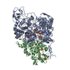
| ||||||||
| 2 | 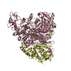
| ||||||||
| 3 | 
| ||||||||
| Unit cell |
| ||||||||
| Details | Dimer confirmed by gel filtration and SAXS |
-Components
-DMSO reductase family type II enzyme, ... , 2 types, 6 molecules ACEBDF
| #1: Protein | Mass: 102069.859 Da / Num. of mol.: 3 / Source method: isolated from a natural source Source: (natural) Azospira oryzae (strain ATCC BAA-33 / DSM 13638 / PS) (bacteria)Strain: ATCC BAA-33 / DSM 13638 / PS / References: UniProt: G8QM55 #2: Protein | Mass: 37067.809 Da / Num. of mol.: 3 / Source method: isolated from a natural source Source: (natural) Azospira oryzae (strain ATCC BAA-33 / DSM 13638 / PS) (bacteria)Strain: ATCC BAA-33 / DSM 13638 / PS / References: UniProt: G8QM54 |
|---|
-Non-polymers , 12 types, 1327 molecules 


| #3: Chemical | ChemComp-SF4 /  Mass: 351.640 Da / Num. of mol.: 12 / Source method: obtained synthetically / Formula: Fe4S4 Mass: 351.640 Da / Num. of mol.: 12 / Source method: obtained synthetically / Formula: Fe4S4#4: Chemical |  Mass: 95.940 Da / Num. of mol.: 3 / Source method: obtained synthetically / Formula: Mo Mass: 95.940 Da / Num. of mol.: 3 / Source method: obtained synthetically / Formula: Mo#5: Chemical |  Mass: 740.557 Da / Num. of mol.: 3 / Source method: obtained synthetically / Formula: C20H26N10O13P2S2 Mass: 740.557 Da / Num. of mol.: 3 / Source method: obtained synthetically / Formula: C20H26N10O13P2S2#6: Chemical |  Mass: 740.557 Da / Num. of mol.: 3 / Source method: obtained synthetically / Formula: C20H26N10O13P2S2 Mass: 740.557 Da / Num. of mol.: 3 / Source method: obtained synthetically / Formula: C20H26N10O13P2S2#7: Chemical | ChemComp-EDO /  Mass: 62.068 Da / Num. of mol.: 24 / Source method: obtained synthetically / Formula: C2H6O2 Mass: 62.068 Da / Num. of mol.: 24 / Source method: obtained synthetically / Formula: C2H6O2#8: Chemical |  Mass: 127.966 Da / Num. of mol.: 3 / Source method: obtained synthetically / Formula: HO3Se Mass: 127.966 Da / Num. of mol.: 3 / Source method: obtained synthetically / Formula: HO3Se#9: Chemical |  Mass: 80.063 Da / Num. of mol.: 3 / Source method: obtained synthetically / Formula: SO3 Mass: 80.063 Da / Num. of mol.: 3 / Source method: obtained synthetically / Formula: SO3#10: Chemical | ChemComp-NA /  Mass: 22.990 Da / Num. of mol.: 8 / Source method: obtained synthetically / Formula: Na Mass: 22.990 Da / Num. of mol.: 8 / Source method: obtained synthetically / Formula: Na#11: Chemical |  Mass: 65.409 Da / Num. of mol.: 3 / Source method: obtained synthetically / Formula: Zn Mass: 65.409 Da / Num. of mol.: 3 / Source method: obtained synthetically / Formula: Zn#12: Chemical |  Mass: 295.795 Da / Num. of mol.: 3 / Source method: obtained synthetically / Formula: Fe3S4 Mass: 295.795 Da / Num. of mol.: 3 / Source method: obtained synthetically / Formula: Fe3S4#13: Chemical | ChemComp-GOL / |  Mass: 92.094 Da / Num. of mol.: 1 / Source method: obtained synthetically / Formula: C3H8O3 Mass: 92.094 Da / Num. of mol.: 1 / Source method: obtained synthetically / Formula: C3H8O3#14: Water | ChemComp-HOH / | Mass: 18.015 Da / Num. of mol.: 1261 / Source method: isolated from a natural source / Formula: H2O |
|---|
-Experimental details
-Experiment
| Experiment | Method: X-RAY DIFFRACTION |
|---|
- Sample preparation
| Crystal | Density Matthews: 2.73 Å3/Da / Density % sol: 55.02 % / Description: Rod |
|---|---|
| Crystal grow | Temperature: 293 K / Method: vapor diffusion, hanging drop / pH: 8 / Details: 20% PEG6K, 0.1 M Tris pH 8.5 / PH range: 8-8.5 |
-Data collection
| Diffraction | Mean temperature: 100 K |
|---|---|
| Diffraction source | Source: SYNCHROTRON / Site: ALS  / Beamline: 12.3.1 / Wavelength: 0.9793 Å / Beamline: 12.3.1 / Wavelength: 0.9793 Å |
| Detector | Type: ADSC QUANTUM 315r / Detector: CCD / Date: Apr 19, 2015 |
| Radiation | Protocol: SINGLE WAVELENGTH / Monochromatic (M) / Laue (L): M / Scattering type: x-ray |
| Radiation wavelength | Wavelength: 0.9793 Å / Relative weight: 1 |
| Reflection | Resolution: 2.38→48.52 Å / Num. obs: 183135 / % possible obs: 100 % / Redundancy: 14.9 % / Rmerge(I) obs: 0.22 / Net I/σ(I): 13 |
| Reflection shell | Resolution: 2.38→2.42 Å / Redundancy: 15 % / Mean I/σ(I) obs: 2 / % possible all: 100 |
- Processing
| Software |
| |||||||||||||||||||||||||||||||||||||||||||||||||||||||||||||||||||||||||||||||||||||||||||||||||||||||||||||||||||||||||||||||||||||||||||||||||||||||||||||||||||||||||||||||||||||||||||||||||||||||||||||||||||||||||
|---|---|---|---|---|---|---|---|---|---|---|---|---|---|---|---|---|---|---|---|---|---|---|---|---|---|---|---|---|---|---|---|---|---|---|---|---|---|---|---|---|---|---|---|---|---|---|---|---|---|---|---|---|---|---|---|---|---|---|---|---|---|---|---|---|---|---|---|---|---|---|---|---|---|---|---|---|---|---|---|---|---|---|---|---|---|---|---|---|---|---|---|---|---|---|---|---|---|---|---|---|---|---|---|---|---|---|---|---|---|---|---|---|---|---|---|---|---|---|---|---|---|---|---|---|---|---|---|---|---|---|---|---|---|---|---|---|---|---|---|---|---|---|---|---|---|---|---|---|---|---|---|---|---|---|---|---|---|---|---|---|---|---|---|---|---|---|---|---|---|---|---|---|---|---|---|---|---|---|---|---|---|---|---|---|---|---|---|---|---|---|---|---|---|---|---|---|---|---|---|---|---|---|---|---|---|---|---|---|---|---|---|---|---|---|---|---|---|---|
| Refinement | Method to determine structure: MOLECULAR REPLACEMENT Starting model: 4YDD Resolution: 2.38→48.52 Å / SU ML: 0.31 / Cross valid method: FREE R-VALUE / σ(F): 1.34 / Phase error: 24.02 / Stereochemistry target values: ML
| |||||||||||||||||||||||||||||||||||||||||||||||||||||||||||||||||||||||||||||||||||||||||||||||||||||||||||||||||||||||||||||||||||||||||||||||||||||||||||||||||||||||||||||||||||||||||||||||||||||||||||||||||||||||||
| Solvent computation | Shrinkage radii: 0.9 Å / VDW probe radii: 1.11 Å / Solvent model: FLAT BULK SOLVENT MODEL | |||||||||||||||||||||||||||||||||||||||||||||||||||||||||||||||||||||||||||||||||||||||||||||||||||||||||||||||||||||||||||||||||||||||||||||||||||||||||||||||||||||||||||||||||||||||||||||||||||||||||||||||||||||||||
| Refinement step | Cycle: LAST / Resolution: 2.38→48.52 Å
| |||||||||||||||||||||||||||||||||||||||||||||||||||||||||||||||||||||||||||||||||||||||||||||||||||||||||||||||||||||||||||||||||||||||||||||||||||||||||||||||||||||||||||||||||||||||||||||||||||||||||||||||||||||||||
| Refine LS restraints |
| |||||||||||||||||||||||||||||||||||||||||||||||||||||||||||||||||||||||||||||||||||||||||||||||||||||||||||||||||||||||||||||||||||||||||||||||||||||||||||||||||||||||||||||||||||||||||||||||||||||||||||||||||||||||||
| LS refinement shell |
|
