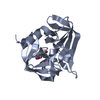
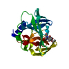
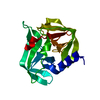
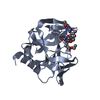
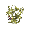
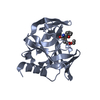
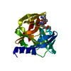
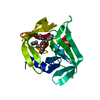
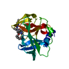
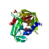
Entry Database : PDB / ID : 5c20Title Crystal structure of EV71 3C Proteinase in complex with Compound 2 3C proteinase Keywords / / / / Function / homology Function Domain/homology Component
/ / / / / / / / / / / / / / / / / / / / / / / / / / / / / / / / / / / / / / / / / / / / / / / / / / / / / / / / / / / / / / / / / / / / / / / / / / / / / / / / / Biological species Method / / Resolution : 2.75 Å Authors Zhang, L. / Huang, G. / Cai, Q. / Zhao, C. / Ren, H. / Li, P. / Li, N. / Chen, S. / Li, J. / Lin, T. Journal : J.Mol.Recognit. / Year : 2016Title : Optimize the interactions at S4 with efficient inhibitors targeting 3C proteinase from enterovirus 71Authors : Zhang, L. / Huang, G. / Cai, Q. / Zhao, C. / Tang, L. / Ren, H. / Li, P. / Li, N. / Huang, J. / Chen, X. / Guan, Y. / You, H. / Chen, S. / Li, J. / Lin, T. History Deposition Jun 15, 2015 Deposition site / Processing site Revision 1.0 Jun 1, 2016 Provider / Type Revision 1.1 Oct 26, 2016 Group Revision 1.2 Sep 27, 2017 Group / Derived calculations / Category / pdbx_struct_oper_listItem / _pdbx_struct_oper_list.symmetry_operationRevision 2.0 Sep 2, 2020 Group / Non-polymer description / Structure summaryCategory / entity / pdbx_entity_nonpolyItem _chem_comp.formula / _chem_comp.formula_weight ... _chem_comp.formula / _chem_comp.formula_weight / _chem_comp.name / _entity.formula_weight / _entity.pdbx_description / _pdbx_entity_nonpoly.name Revision 2.1 Nov 8, 2023 Group / Database references / Refinement descriptionCategory chem_comp_atom / chem_comp_bond ... chem_comp_atom / chem_comp_bond / database_2 / pdbx_initial_refinement_model Item / _database_2.pdbx_database_accessionRevision 2.2 Oct 9, 2024 Group / Category / pdbx_modification_feature
Show all Show less
 Movie
Movie Controller

 Yorodumi
 Open data
 Basic information
 Components
 Keywords
 Function and homology information
 Enterovirus A71
 X-RAY DIFFRACTION /
  Authors
 Citation
 Structure visualization
 Downloads & links
 Other downloads
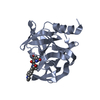


 PDBj
PDBj

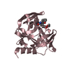
 Assembly
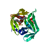

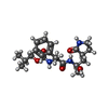
 Mass: 405.488 Da / Num. of mol.: 1 / Source method: obtained synthetically / Formula: C21H31N3O5
 Mass: 18.015 Da / Num. of mol.: 13 / Source method: isolated from a natural source / Formula: H2O
 Sample preparation
 Processing