 Movie
Movie Controller


+ Open data

- Basic information
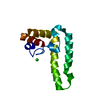
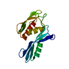
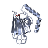
| Entry | Database: PDB / ID: 5b79 | ||||||
|---|---|---|---|---|---|---|---|
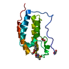
| Title | Crystal structure of DPF2 double PHD finger | ||||||
 Components Components | Zinc finger protein ubi-d4 | ||||||
 Keywords Keywords | METAL BINDING PROTEIN / DPF2 | ||||||
| Function / homology |  Function and homology information Function and homology informationnegative regulation of myeloid progenitor cell differentiation / Formation of the embryonic stem cell BAF (esBAF) complex / nBAF complex / Formation of the canonical BAF (cBAF) complex / Formation of neuronal progenitor and neuronal BAF (npBAF and nBAF) / regulation of G0 to G1 transition / histone H3K14ac reader activity / regulation of nucleotide-excision repair / regulation of mitotic metaphase/anaphase transition / SWI/SNF complex ...negative regulation of myeloid progenitor cell differentiation / Formation of the embryonic stem cell BAF (esBAF) complex / nBAF complex / Formation of the canonical BAF (cBAF) complex / Formation of neuronal progenitor and neuronal BAF (npBAF and nBAF) / regulation of G0 to G1 transition / histone H3K14ac reader activity / regulation of nucleotide-excision repair / regulation of mitotic metaphase/anaphase transition / SWI/SNF complex / positive regulation of double-strand break repair / positive regulation of stem cell population maintenance / Regulation of MITF-M-dependent genes involved in pigmentation / regulation of G1/S transition of mitotic cell cycle / histone H4K16ac reader activity / Regulation of endogenous retroelements by Piwi-interacting RNAs (piRNAs) / apoptotic signaling pathway / transcription corepressor activity / nervous system development / chromatin remodeling / apoptotic process / regulation of transcription by RNA polymerase II / centrosome / chromatin / negative regulation of transcription by RNA polymerase II / zinc ion binding / nucleoplasm / nucleus / cytosol Similarity search - Function | ||||||
| Biological species |  Homo sapiens (human) Homo sapiens (human) | ||||||
| Method |  X-RAY DIFFRACTION / MOLECULAR REPLACEMENT / Resolution: 2.601 Å X-RAY DIFFRACTION / MOLECULAR REPLACEMENT / Resolution: 2.601 Å | ||||||
 Authors Authors | Li, H. / Xiong, X. | ||||||
 Citation Citation | Journal: Nat.Chem.Biol. / Year: 2016 Title: Selective recognition of histone crotonylation by double PHD fingers of MOZ and DPF2 Authors: Xiong, X. / Panchenko, T. / Yang, S. / Zhao, S. / Yan, P. / Zhang, W. / Xie, W. / Li, Y. / Zhao, Y. / Allis, C.D. / Li, H. | ||||||
| History |
|
- Structure visualization
| Structure viewer | Molecule: MolmilJmol/JSmol |
|---|
- Downloads & links
-Download
| PDBx/mmCIF format | 5b79.cif.gz | 37.9 KB | Display | PDBx/mmCIF format |
|---|---|---|---|---|
| PDB format | pdb5b79.ent.gz | 23.9 KB | Display | PDB format |
| PDBx/mmJSON format | 5b79.json.gz | Tree view | PDBx/mmJSON format | |
| Others |  Other downloads Other downloads |
-Validation report
| Arichive directory | https://data.pdbj.org/pub/pdb/validation_reports/b7/5b79ftp://data.pdbj.org/pub/pdb/validation_reports/b7/5b79 | HTTPS FTP |
|---|
-Related structure data
| Related structure data | 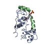 5b75C 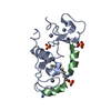 5b76C 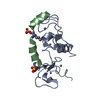 5b77C 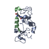 5b78C 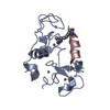 4llbS S: Starting model for refinement C: citing same article ( |
|---|---|
| Similar structure data |


-Links
 PDBj
PDBj


- Assembly
| Deposited unit | 
| ||||||||
|---|---|---|---|---|---|---|---|---|---|
| 1 |
| ||||||||
| Unit cell |
|
-Components
| #1: Protein | Mass: 13818.602 Da / Num. of mol.: 1 / Fragment: UNP residues 270-391 Source method: isolated from a genetically manipulated source Source: (gene. exp.) Homo sapiens (human) / Gene: DPF2 / Production host:  | ||
|---|---|---|---|
| #2: Chemical | ChemComp-ZN /   Mass: 65.409 Da / Num. of mol.: 4 / Source method: obtained synthetically / Formula: Zn Mass: 65.409 Da / Num. of mol.: 4 / Source method: obtained synthetically / Formula: Zn#3: Water | ChemComp-HOH / |  Mass: 18.015 Da / Num. of mol.: 10 / Source method: isolated from a natural source / Formula: H2O Mass: 18.015 Da / Num. of mol.: 10 / Source method: isolated from a natural source / Formula: H2O |
-Experimental details
-Experiment
| Experiment | Method: X-RAY DIFFRACTION / Number of used crystals: 1 |
|---|
- Sample preparation
| Crystal | Density Matthews: 2.72 Å3/Da / Density % sol: 54.82 % |
|---|---|
| Crystal grow | Temperature: 291 K / Method: vapor diffusion, sitting drop / Details: ammonium citrate tribasic, Bis-Tris propane |
-Data collection
| Diffraction | Mean temperature: 100 K |
|---|---|
| Diffraction source | Source: ROTATING ANODE / Type: RIGAKU MICROMAX-007 HF / Wavelength: 1.5418 Å |
| Detector | Type: DECTRIS PILATUS 200K / Detector: PIXEL / Date: Feb 5, 2015 |
| Radiation | Protocol: SINGLE WAVELENGTH / Monochromatic (M) / Laue (L): M / Scattering type: x-ray |
| Radiation wavelength | Wavelength: 1.5418 Å / Relative weight: 1 |
| Reflection | Resolution: 2.6→23.6 Å / Num. obs: 4902 / % possible obs: 99.6 % / Redundancy: 7.5 % / Net I/σ(I): 29.9 |
| Reflection shell | Resolution: 2.6→2.64 Å |
- Processing
| Software |
| ||||||||||||||||||||||||||||
|---|---|---|---|---|---|---|---|---|---|---|---|---|---|---|---|---|---|---|---|---|---|---|---|---|---|---|---|---|---|
| Refinement | Method to determine structure: MOLECULAR REPLACEMENT Starting model: 4LLB Resolution: 2.601→23.599 Å / SU ML: 0.23 / Cross valid method: FREE R-VALUE / σ(F): 1.35 / Phase error: 24.68
| ||||||||||||||||||||||||||||
| Solvent computation | Shrinkage radii: 0.9 Å / VDW probe radii: 1.11 Å | ||||||||||||||||||||||||||||
| Refinement step | Cycle: LAST / Resolution: 2.601→23.599 Å
| ||||||||||||||||||||||||||||
| Refine LS restraints |
| ||||||||||||||||||||||||||||
| LS refinement shell |
|
