 Movie
Movie Controller

[English] 日本語
 Yorodumi
- PDB-5b6v: A three dimensional movie of structural changes in bacteriorhodop... -
+ Open data

- Basic information
| Entry | Database: PDB / ID: 5b6v | ||||||
|---|---|---|---|---|---|---|---|
| Title | A three dimensional movie of structural changes in bacteriorhodopsin: resting state structure | ||||||
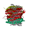
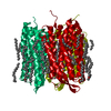
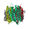
 Components Components | Bacteriorhodopsin | ||||||
 Keywords Keywords | PROTON TRANSPORT / bacteriorhodopsin / XFEL / time-resolved SFX / SACLA | ||||||
| Function / homology |  Function and homology information Function and homology informationlight-driven active monoatomic ion transmembrane transporter activity / photoreceptor activity / phototransduction / monoatomic ion channel activity / proton transmembrane transport / plasma membrane Similarity search - Function | ||||||
| Biological species |  Halobacterium salinarum (Halophile) Halobacterium salinarum (Halophile) | ||||||
| Method |  X-RAY DIFFRACTION / FREE ELECTRON LASER / MOLECULAR REPLACEMENT / Resolution: 2 Å X-RAY DIFFRACTION / FREE ELECTRON LASER / MOLECULAR REPLACEMENT / Resolution: 2 Å | ||||||
 Authors Authors | Nango, E. / Royant, A. / Nakane, T. / Tanaka, T. / Arima, T. / Neutze, R. / Iwata, S. | ||||||
 Citation Citation | Journal: Science / Year: 2016 Title: A three-dimensional movie of structural changes in bacteriorhodopsin Authors: Nango, E. / Royant, A. / Kubo, M. / Nakane, T. / Wickstrand, C. / Kimura, T. / Tanaka, T. / Tono, K. / Song, C. / Tanaka, R. / Arima, T. / Yamashita, A. / Kobayashi, J. / Hosaka, T. / ...Authors: Nango, E. / Royant, A. / Kubo, M. / Nakane, T. / Wickstrand, C. / Kimura, T. / Tanaka, T. / Tono, K. / Song, C. / Tanaka, R. / Arima, T. / Yamashita, A. / Kobayashi, J. / Hosaka, T. / Mizohata, E. / Nogly, P. / Sugahara, M. / Nam, D. / Nomura, T. / Shimamura, T. / Im, D. / Fujiwara, T. / Yamanaka, Y. / Jeon, B. / Nishizawa, T. / Oda, K. / Fukuda, M. / Andersson, R. / Bath, P. / Dods, R. / Davidsson, J. / Matsuoka, S. / Kawatake, S. / Murata, M. / Nureki, O. / Owada, S. / Kameshima, T. / Hatsui, T. / Joti, Y. / Schertler, G. / Yabashi, M. / Bondar, A.N. / Standfuss, J. / Neutze, R. / Iwata, S. | ||||||
| History |
|
- Structure visualization
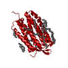
| Structure viewer | Molecule: MolmilJmol/JSmol |
|---|
- Downloads & links
-Download
| PDBx/mmCIF format | 5b6v.cif.gz | 119.3 KB | Display | PDBx/mmCIF format |
|---|---|---|---|---|
| PDB format | pdb5b6v.ent.gz | 93 KB | Display | PDB format |
| PDBx/mmJSON format | 5b6v.json.gz | Tree view | PDBx/mmJSON format | |
| Others |  Other downloads Other downloads |
-Validation report
| Arichive directory | https://data.pdbj.org/pub/pdb/validation_reports/b6/5b6vftp://data.pdbj.org/pub/pdb/validation_reports/b6/5b6v | HTTPS FTP |
|---|
-Related structure data
| Related structure data |  5b6wC 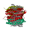 5b6xC 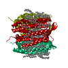 5b6yC 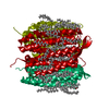 5b6zC 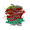 5h2hC  5h2iC 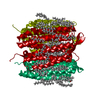 5h2jC 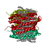 5h2kC 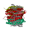 5h2lC  5h2mC  5h2nC  5h2oC 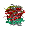 5h2pC  3ns0S C: citing same article ( S: Starting model for refinement |
|---|---|
| Similar structure data | |
| Experimental dataset #1 | Data reference: 10.11577/1337005 / Data set type: diffraction image data |


-Links
 PDBj
PDBj


- Assembly
| Deposited unit | 
| ||||||||
|---|---|---|---|---|---|---|---|---|---|
| 1 | 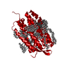
| ||||||||
| Unit cell |
|
-Components
-Protein , 1 types, 1 molecules A
| #1: Protein | Mass: 26814.412 Da / Num. of mol.: 1 / Fragment: UNP residues 14-261 / Source method: isolated from a natural source Source: (natural) Halobacterium salinarum (strain ATCC 700922 / JCM 11081 / NRC-1) (Halophile)Strain: ATCC 700922 / JCM 11081 / NRC-1 / References: UniProt: P02945 |
|---|
-Non-polymers , 11 types, 33 molecules 
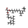


| #2: Chemical | ChemComp-RET /  Mass: 284.436 Da / Num. of mol.: 1 / Source method: obtained synthetically / Formula: C20H28O Mass: 284.436 Da / Num. of mol.: 1 / Source method: obtained synthetically / Formula: C20H28O | ||||||||||||||||||
|---|---|---|---|---|---|---|---|---|---|---|---|---|---|---|---|---|---|---|---|
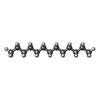
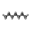
| #3: Chemical |  Mass: 653.157 Da / Num. of mol.: 3 / Source method: obtained synthetically / Formula: C43H88O3 Mass: 653.157 Da / Num. of mol.: 3 / Source method: obtained synthetically / Formula: C43H88O3#4: Chemical | ChemComp-TRD / |  Mass: 184.361 Da / Num. of mol.: 1 / Source method: obtained synthetically / Formula: C13H28 Mass: 184.361 Da / Num. of mol.: 1 / Source method: obtained synthetically / Formula: C13H28#5: Chemical | ChemComp-D10 / |  Mass: 142.282 Da / Num. of mol.: 1 / Source method: obtained synthetically / Formula: C10H22 Mass: 142.282 Da / Num. of mol.: 1 / Source method: obtained synthetically / Formula: C10H22#6: Chemical | ChemComp-HP6 / |  Mass: 100.202 Da / Num. of mol.: 1 / Source method: obtained synthetically / Formula: C7H16 Mass: 100.202 Da / Num. of mol.: 1 / Source method: obtained synthetically / Formula: C7H16#7: Chemical |  Mass: 114.229 Da / Num. of mol.: 3 / Source method: obtained synthetically / Formula: C8H18 Mass: 114.229 Da / Num. of mol.: 3 / Source method: obtained synthetically / Formula: C8H18#8: Chemical |  Mass: 212.415 Da / Num. of mol.: 2 / Source method: obtained synthetically / Formula: C15H32 Mass: 212.415 Da / Num. of mol.: 2 / Source method: obtained synthetically / Formula: C15H32#9: Chemical |  Mass: 156.308 Da / Num. of mol.: 2 / Source method: obtained synthetically / Formula: C11H24 Mass: 156.308 Da / Num. of mol.: 2 / Source method: obtained synthetically / Formula: C11H24#10: Chemical | ChemComp-DD9 / |  Mass: 128.255 Da / Num. of mol.: 1 / Source method: obtained synthetically / Formula: C9H20 Mass: 128.255 Da / Num. of mol.: 1 / Source method: obtained synthetically / Formula: C9H20#11: Chemical | ChemComp-C14 / |  Mass: 198.388 Da / Num. of mol.: 1 / Source method: obtained synthetically / Formula: C14H30 Mass: 198.388 Da / Num. of mol.: 1 / Source method: obtained synthetically / Formula: C14H30#12: Water | ChemComp-HOH / | Mass: 18.015 Da / Num. of mol.: 17 / Source method: isolated from a natural source / Formula: H2O |
-Details
| Has protein modification | Y |
|---|---|
| Nonpolymer details | Ligands TRD, D10, HP6, OCT, MYS, UND, DD9, and C14 are modeled as a lipid fragment. |
-Experimental details
-Experiment
| Experiment | Method: X-RAY DIFFRACTION / Number of used crystals: 1 |
|---|
- Sample preparation
| Crystal | Density Matthews: 2.35 Å3/Da / Density % sol: 47.77 % |
|---|---|
| Crystal grow | Temperature: 293 K / Method: lipidic cubic phase / Details: 0.1M Na/K phosphate pH5.4 , 30% PEG 2000 |
-Data collection
| Diffraction | Mean temperature: 293 K |
|---|---|
| Diffraction source | Source: FREE ELECTRON LASER / Site: SACLA  / Beamline: BL3 / Wavelength: 1.63 Å / Beamline: BL3 / Wavelength: 1.63 Å |
| Detector | Type: MPCCD / Detector: CCD / Date: Feb 4, 2016 |
| Radiation | Protocol: SINGLE WAVELENGTH / Monochromatic (M) / Laue (L): M / Scattering type: x-ray |
| Radiation wavelength | Wavelength: 1.63 Å / Relative weight: 1 |
| Reflection | Resolution: 2→39.92 Å / Num. obs: 16786 / % possible obs: 100 % / Redundancy: 3389 % / Net I/σ(I): 30 |
| Reflection shell | Resolution: 2→2.07 Å |
- Processing
| Software |
| |||||||||||||||||||||||||||||||||||||||||||||||||
|---|---|---|---|---|---|---|---|---|---|---|---|---|---|---|---|---|---|---|---|---|---|---|---|---|---|---|---|---|---|---|---|---|---|---|---|---|---|---|---|---|---|---|---|---|---|---|---|---|---|---|
| Refinement | Method to determine structure: MOLECULAR REPLACEMENT Starting model: 3NS0 Resolution: 2→21.912 Å / SU ML: 0.22 / Cross valid method: FREE R-VALUE / σ(F): 1.38 / Phase error: 19.65
| |||||||||||||||||||||||||||||||||||||||||||||||||
| Solvent computation | Shrinkage radii: 0.9 Å / VDW probe radii: 1.11 Å | |||||||||||||||||||||||||||||||||||||||||||||||||
| Refinement step | Cycle: LAST / Resolution: 2→21.912 Å
| |||||||||||||||||||||||||||||||||||||||||||||||||
| Refine LS restraints |
| |||||||||||||||||||||||||||||||||||||||||||||||||
| LS refinement shell |
|
