| 登録情報 | データベース: PDB / ID: 5awt
|
|---|
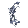
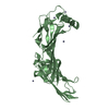
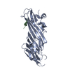
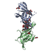
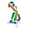
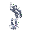
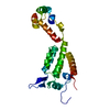
| タイトル | Crystal structure of the SGIP1 mu homology domain in complex with an Eps15 fragment containing two DPF motifs (YDPFGGDPFKG) |
|---|
 要素 要素 | - Epidermal growth factor receptor substrate 15
- SH3-containing GRB2-like protein 3-interacting protein 1
|
|---|
 キーワード キーワード | ENDOCYTOSIS / Protein-protein interaction |
|---|
| 機能・相同性 |  機能・相同性情報 機能・相同性情報
positive regulation of feeding behavior / ubiquitin-dependent endocytosis / Golgi to endosome transport / clathrin coat of coated pit / AP-2 adaptor complex / postsynaptic endocytic zone / postsynaptic neurotransmitter receptor internalization / vesicle organization / clathrin coat assembly / clathrin-dependent endocytosis ...positive regulation of feeding behavior / ubiquitin-dependent endocytosis / Golgi to endosome transport / clathrin coat of coated pit / AP-2 adaptor complex / postsynaptic endocytic zone / postsynaptic neurotransmitter receptor internalization / vesicle organization / clathrin coat assembly / clathrin-dependent endocytosis / endocytic recycling / clathrin-coated vesicle / aggresome / endosomal transport / ciliary membrane / positive regulation of receptor recycling / response to dietary excess / energy homeostasis / synaptic vesicle endocytosis / polyubiquitin modification-dependent protein binding / InlB-mediated entry of Listeria monocytogenes into host cell / clathrin-coated pit / receptor-mediated endocytosis of virus by host cell / basal plasma membrane / EGFR downregulation / ubiquitin binding / Negative regulation of MET activity / phospholipid binding / positive regulation of receptor-mediated endocytosis / SH3 domain binding / endocytosis / Cargo recognition for clathrin-mediated endocytosis / Clathrin-mediated endocytosis / presynapse / regulation of cell population proliferation / regulation of protein localization / early endosome membrane / microtubule binding / apical plasma membrane / cadherin binding / intracellular membrane-bounded organelle / calcium ion binding / symbiont entry into host cell / glutamatergic synapse / identical protein binding / membrane / plasma membrane / cytosol / cytoplasm類似検索 - 分子機能 SGIP1, mu-homology domain / Muniscin C-terminal / Muniscin C-terminal mu homology domain / EH domain / EH domain profile. / Eps15 homology domain / EH domain / AP-2 complex subunit mu, C-terminal superfamily / Mu homology domain / Mu homology domain (MHD) profile. ...SGIP1, mu-homology domain / Muniscin C-terminal / Muniscin C-terminal mu homology domain / EH domain / EH domain profile. / Eps15 homology domain / EH domain / AP-2 complex subunit mu, C-terminal superfamily / Mu homology domain / Mu homology domain (MHD) profile. / Ubiquitin-interacting motif. / Ubiquitin interacting motif / Ubiquitin-interacting motif (UIM) domain profile. / EF-hand, calcium binding motif / EF-Hand 1, calcium-binding site / EF-hand calcium-binding domain. / EF-hand calcium-binding domain profile. / EF-hand domain / EF-hand domain pair類似検索 - ドメイン・相同性 Epidermal growth factor receptor substrate 15 / SH3-containing GRB2-like protein 3-interacting protein 1類似検索 - 構成要素 |
|---|
| 生物種 |  Homo sapiens (ヒト) Homo sapiens (ヒト) |
|---|
| 手法 |  X線回折 / シンクロトロン / 分子置換 / 解像度: 2.702 Å X線回折 / シンクロトロン / 分子置換 / 解像度: 2.702 Å |
|---|
 データ登録者 データ登録者 | Shimada, A. / Yamaguchi, A. / Kohda, D. |
|---|
| 資金援助 |  日本, 3件 日本, 3件 | 組織 | 認可番号 | 国 |
|---|
| MEXT/JSPS | KAKENHI 20687006 | 日本 | | MEXT/JSPS | KAKENHI 24687014 | 日本 | | MEXT/JSPS | KAKENHI 25121726 | 日本 |
|
|---|
 引用 引用 | ジャーナル: Sci Rep / 年: 2016
タイトル: Structural basis for the recognition of two consecutive mutually interacting DPF motifs by the SGIP1 mu homology domain.
著者: Shimada, A. / Yamaguchi, A. / Kohda, D. |
|---|
| 履歴 | | 登録 | 2015年7月8日 | 登録サイト: PDBJ / 処理サイト: PDBJ |
|---|
| 改定 1.0 | 2016年7月6日 | Provider: repository / タイプ: Initial release |
|---|
| 改定 1.1 | 2020年2月26日 | Group: Data collection / Derived calculations / カテゴリ: diffrn_source / pdbx_struct_oper_list
Item: _diffrn_source.pdbx_synchrotron_site / _pdbx_struct_oper_list.symmetry_operation |
|---|
| 改定 1.2 | 2023年11月8日 | Group: Data collection / Database references ...Data collection / Database references / Derived calculations / Refinement description
カテゴリ: chem_comp_atom / chem_comp_bond ...chem_comp_atom / chem_comp_bond / database_2 / pdbx_initial_refinement_model / struct_conn
Item: _database_2.pdbx_DOI / _database_2.pdbx_database_accession ..._database_2.pdbx_DOI / _database_2.pdbx_database_accession / _struct_conn.pdbx_dist_value / _struct_conn.ptnr1_auth_comp_id / _struct_conn.ptnr1_auth_seq_id / _struct_conn.ptnr1_label_asym_id / _struct_conn.ptnr1_label_atom_id / _struct_conn.ptnr1_label_comp_id / _struct_conn.ptnr1_label_seq_id / _struct_conn.ptnr2_auth_comp_id / _struct_conn.ptnr2_auth_seq_id / _struct_conn.ptnr2_label_asym_id / _struct_conn.ptnr2_label_atom_id / _struct_conn.ptnr2_label_comp_id / _struct_conn.ptnr2_symmetry |
|---|
| 改定 1.3 | 2024年11月6日 | Group: Structure summary
カテゴリ: pdbx_entry_details / pdbx_modification_feature |
|---|
|
|---|
 ムービー
ムービー コントローラー


 データを開く
 基本情報
基本情報 構造の表示
構造の表示 ダウンロードとリンク
 その他のダウンロード


 PDBj
PDBj


 集合体
集合体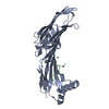


 分子量: 65.409 Da / 分子数: 6 / 由来タイプ: 合成 / 式: Zn
 分子量: 18.015 Da / 分子数: 35 / 由来タイプ: 天然 / 式: H2O
 試料調製
試料調製 解析
解析