 Movie
Movie Controller

[English] 日本語
 Yorodumi
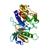
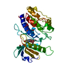
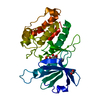
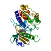
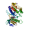
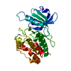
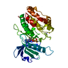
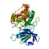
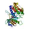
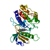
- PDB-5av0: Crystal structure of DAPK1 in complex with 7,3',4'-trihydroxyisof... -
+ Open data

- Basic information
| Entry | Database: PDB / ID: 5av0 | ||||||
|---|---|---|---|---|---|---|---|
| Title | Crystal structure of DAPK1 in complex with 7,3',4'-trihydroxyisoflavone. | ||||||
 Components Components | Death-associated protein kinase 1 | ||||||
 Keywords Keywords | TRANSFERASE / death-associated protein kinase 1 / serine/threonine protein kinase / natural flavonoid | ||||||
| Function / homology |  Function and homology information Function and homology informationcellular response to hydroperoxide / negative regulation of kinase activity / regulation of response to tumor cell / positive regulation of autophagic cell death / Caspase activation via Dependence Receptors in the absence of ligand / defense response to tumor cell / calcium/calmodulin-dependent protein kinase activity / regulation of NMDA receptor activity / syntaxin-1 binding / positive regulation of autophagy ...cellular response to hydroperoxide / negative regulation of kinase activity / regulation of response to tumor cell / positive regulation of autophagic cell death / Caspase activation via Dependence Receptors in the absence of ligand / defense response to tumor cell / calcium/calmodulin-dependent protein kinase activity / regulation of NMDA receptor activity / syntaxin-1 binding / positive regulation of autophagy / regulation of autophagy / apoptotic signaling pathway / cellular response to type II interferon / protein autophosphorylation / actin cytoskeleton / regulation of apoptotic process / protein phosphorylation / protein kinase activity / calmodulin binding / non-specific serine/threonine protein kinase / negative regulation of translation / postsynaptic density / intracellular signal transduction / positive regulation of apoptotic process / protein serine kinase activity / protein serine/threonine kinase activity / apoptotic process / negative regulation of apoptotic process / GTP binding / glutamatergic synapse / ATP binding / identical protein binding / nucleus / plasma membrane / cytoplasm Similarity search - Function | ||||||
| Biological species |  Homo sapiens (human) Homo sapiens (human) | ||||||
| Method |  X-RAY DIFFRACTION / SYNCHROTRON / Resolution: 1.85 Å X-RAY DIFFRACTION / SYNCHROTRON / Resolution: 1.85 Å | ||||||
 Authors Authors | Yokoyama, T. / Mizuguchi, M. | ||||||
 Citation Citation | Journal: J.Med.Chem. / Year: 2015 Title: Structural Insight into the Interactions between Death-Associated Protein Kinase 1 and Natural Flavonoids. Authors: Yokoyama, T. / Kosaka, Y. / Mizuguchi, M. | ||||||
| History |
|
- Structure visualization
| Structure viewer | Molecule: MolmilJmol/JSmol |
|---|
- Downloads & links
-Download
| PDBx/mmCIF format | 5av0.cif.gz | 73.7 KB | Display | PDBx/mmCIF format |
|---|---|---|---|---|
| PDB format | pdb5av0.ent.gz | 53 KB | Display | PDB format |
| PDBx/mmJSON format | 5av0.json.gz | Tree view | PDBx/mmJSON format | |
| Others |  Other downloads Other downloads |
-Validation report
| Arichive directory | https://data.pdbj.org/pub/pdb/validation_reports/av/5av0ftp://data.pdbj.org/pub/pdb/validation_reports/av/5av0 | HTTPS FTP |
|---|
-Related structure data
| Related structure data |  5autC  5auuC  5auvC 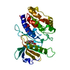 5auwC  5auxC  5auyC  5auzC 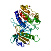 5av1C  5av2C  5av3C  5av4C C: citing same article ( |
|---|---|
| Similar structure data |


-Links
 PDBj
PDBj


- Assembly
| Deposited unit | 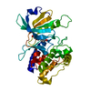
| ||||||||
|---|---|---|---|---|---|---|---|---|---|
| 1 |
| ||||||||
| Unit cell |
|
-Components
| #1: Protein | Mass: 33796.402 Da / Num. of mol.: 1 / Fragment: UNP RESIDUES 1-285 Source method: isolated from a genetically manipulated source Source: (gene. exp.) Homo sapiens (human) / Gene: DAPK1, DAPK / Production host:  References: UniProt: P53355, non-specific serine/threonine protein kinase |
|---|---|
| #2: Chemical | ChemComp-47X / 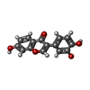  Mass: 270.237 Da / Num. of mol.: 1 / Source method: obtained synthetically / Formula: C15H10O5 Mass: 270.237 Da / Num. of mol.: 1 / Source method: obtained synthetically / Formula: C15H10O5 |
| #3: Water | ChemComp-HOH /  Mass: 18.015 Da / Num. of mol.: 192 / Source method: isolated from a natural source / Formula: H2O Mass: 18.015 Da / Num. of mol.: 192 / Source method: isolated from a natural source / Formula: H2O |
-Experimental details
-Experiment
| Experiment | Method: X-RAY DIFFRACTION |
|---|
- Sample preparation
| Crystal | Density Matthews: 1.89 Å3/Da / Density % sol: 34.95 % |
|---|---|
| Crystal grow | Temperature: 293 K / Method: vapor diffusion, hanging drop / Details: 2 M ammonium sulfate, 0.1 M MES |
-Data collection
| Diffraction | Mean temperature: 100 K |
|---|---|
| Diffraction source | Source: SYNCHROTRON / Site: Photon Factory  / Beamline: BL-5A / Wavelength: 1 Å / Beamline: BL-5A / Wavelength: 1 Å |
| Detector | Type: ADSC QUANTUM 315r / Detector: CCD / Date: Nov 2, 2014 |
| Radiation | Protocol: SINGLE WAVELENGTH / Monochromatic (M) / Laue (L): M / Scattering type: x-ray |
| Radiation wavelength | Wavelength: 1 Å / Relative weight: 1 |
| Reflection | Resolution: 1.85→35.9 Å / Num. obs: 21813 / % possible obs: 96.6 % / Redundancy: 3.8 % / Net I/σ(I): 23.7 |
- Processing
| Software |
| |||||||||||||||||||||||||||||||||||||||||||||||||||||||||||||||
|---|---|---|---|---|---|---|---|---|---|---|---|---|---|---|---|---|---|---|---|---|---|---|---|---|---|---|---|---|---|---|---|---|---|---|---|---|---|---|---|---|---|---|---|---|---|---|---|---|---|---|---|---|---|---|---|---|---|---|---|---|---|---|---|---|
| Refinement | Resolution: 1.85→35.9 Å / SU ML: 0.16 / Cross valid method: FREE R-VALUE / σ(F): 1.38 / Phase error: 20.13 / Stereochemistry target values: ML
| |||||||||||||||||||||||||||||||||||||||||||||||||||||||||||||||
| Solvent computation | Shrinkage radii: 0.9 Å / VDW probe radii: 1.11 Å / Solvent model: FLAT BULK SOLVENT MODEL | |||||||||||||||||||||||||||||||||||||||||||||||||||||||||||||||
| Refinement step | Cycle: LAST / Resolution: 1.85→35.9 Å
| |||||||||||||||||||||||||||||||||||||||||||||||||||||||||||||||
| Refine LS restraints |
| |||||||||||||||||||||||||||||||||||||||||||||||||||||||||||||||
| LS refinement shell |
|
