 Movie
Movie Controller


+ Open data

- Basic information
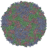
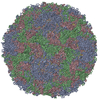
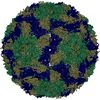
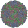
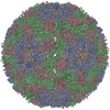
| Entry | Database: PDB / ID: 5aoo | ||||||
|---|---|---|---|---|---|---|---|
| Title | X-ray structure of a human Kobuvirus: Aichi virus A (AiV) | ||||||
 Components Components |
| ||||||
 Keywords Keywords | VIRUS / AICHI VIRUS / KOBUVIRUS / PICORNAVIRUS / ACUTE GASTROENTERISIS / ICOSAHEDRAL CAPSID / FULL VIRUS PARTICLE | ||||||
| Function / homology |  Function and homology information Function and homology informationhost cell Golgi membrane / symbiont-mediated suppression of host mRNA export from nucleus / picornain 3C / T=pseudo3 icosahedral viral capsid / host cell cytoplasmic vesicle membrane / channel activity / monoatomic ion transmembrane transport / RNA helicase activity / RNA helicase / RNA-directed RNA polymerase ...host cell Golgi membrane / symbiont-mediated suppression of host mRNA export from nucleus / picornain 3C / T=pseudo3 icosahedral viral capsid / host cell cytoplasmic vesicle membrane / channel activity / monoatomic ion transmembrane transport / RNA helicase activity / RNA helicase / RNA-directed RNA polymerase / cysteine-type endopeptidase activity / viral RNA genome replication / RNA-directed RNA polymerase activity / symbiont entry into host cell / virion attachment to host cell / DNA-templated transcription / structural molecule activity / ATP hydrolysis activity / proteolysis / RNA binding / ATP binding Similarity search - Function | ||||||
| Biological species |  AICHIVIRUS A AICHIVIRUS A | ||||||
| Method |  X-RAY DIFFRACTION / SYNCHROTRON / MOLECULAR REPLACEMENT / Resolution: 2.1 Å X-RAY DIFFRACTION / SYNCHROTRON / MOLECULAR REPLACEMENT / Resolution: 2.1 Å | ||||||
 Authors Authors | Sabin, C. / Palkova, L. / Plevka, P. | ||||||
 Citation Citation | Journal: Acta Crystallogr.,Sect.F / Year: 2016 Title: The Use of Noncrystallographic Symmetry Averaging to Solve Structures from Data Affected by Perfect Hemihedral Twinning Authors: Sabin, C. / Plevka, P. | ||||||
| History |
|
- Structure visualization
| Structure viewer | Molecule: MolmilJmol/JSmol |
|---|
- Downloads & links
-Download
| PDBx/mmCIF format | 5aoo.cif.gz | 157.7 KB | Display | PDBx/mmCIF format |
|---|---|---|---|---|
| PDB format | pdb5aoo.ent.gz | 123.7 KB | Display | PDB format |
| PDBx/mmJSON format | 5aoo.json.gz | Tree view | PDBx/mmJSON format | |
| Others |  Other downloads Other downloads |
-Validation report
| Arichive directory | https://data.pdbj.org/pub/pdb/validation_reports/ao/5aooftp://data.pdbj.org/pub/pdb/validation_reports/ao/5aoo | HTTPS FTP |
|---|
-Related structure data
| Related structure data | 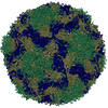 1bevS S: Starting model for refinement |
|---|---|
| Similar structure data |


-Links
 PDBj
PDBj

- Assembly
| Deposited unit | 
| |||||||||||||||
|---|---|---|---|---|---|---|---|---|---|---|---|---|---|---|---|---|
| 1 | x 60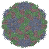
| |||||||||||||||
| 2 |
| |||||||||||||||
| 3 | x 5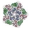
| |||||||||||||||
| 4 | x 6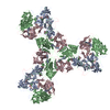
| |||||||||||||||
| 5 | 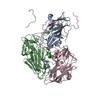
| |||||||||||||||
| Unit cell |
| |||||||||||||||
| Symmetry | Point symmetry: (Schoenflies symbol: I (icosahedral)) | |||||||||||||||
| Noncrystallographic symmetry (NCS) | NCS oper:
|
-Components
| #1: Protein | Mass: 27194.688 Da / Num. of mol.: 1 / Source method: isolated from a natural source / Source: (natural) AICHIVIRUS A / References: UniProt: Q91QP4, UniProt: O91464*PLUS |
|---|---|
| #2: Protein | Mass: 38950.758 Da / Num. of mol.: 1 / Source method: isolated from a natural source / Source: (natural) AICHIVIRUS A / References: UniProt: Q91QP4, UniProt: O91464*PLUS |
| #3: Protein | Mass: 24082.244 Da / Num. of mol.: 1 / Source method: isolated from a natural source / Source: (natural) AICHIVIRUS A / References: UniProt: Q91QP4, UniProt: O91464*PLUS |
| #4: Water | ChemComp-HOH /  Mass: 18.015 Da / Num. of mol.: 147 / Source method: isolated from a natural source / Formula: H2O Mass: 18.015 Da / Num. of mol.: 147 / Source method: isolated from a natural source / Formula: H2O |
-Experimental details
-Experiment
| Experiment | Method: X-RAY DIFFRACTION / Number of used crystals: 1 |
|---|
- Sample preparation
| Crystal | Density Matthews: 4.33 Å3/Da / Density % sol: 71.78 % Description: FOR TWINNING ANALYSES AND CALCULATION OF INITIAL ROTATION FUNCTIONS THE AIV DIFFRACTION IMAGES WERE PROCESSED IN SPACEGROUP I23 TO 2.3 ANGSTROM RESOLUTION IN PROGRAM XDS. HOWEVER, SINCE ...Description: FOR TWINNING ANALYSES AND CALCULATION OF INITIAL ROTATION FUNCTIONS THE AIV DIFFRACTION IMAGES WERE PROCESSED IN SPACEGROUP I23 TO 2.3 ANGSTROM RESOLUTION IN PROGRAM XDS. HOWEVER, SINCE THE DATA ANALYSIS REVEALED THAT THE CRYSTAL WAS PERFECTLY HEMIHEDRALLY TWINNED THE IMAGES WERE RE-PROCESSED IN SPACE GROUP I432. TAKING ADVANTAGE OF THE HIGHER SYMMETRY ALLOWED US TO OBTAIN DATASET THAT WAS MORE THAN 90 PERCENT COMPLETE FROM FIRST 160 DIFFRACTION IMAGES, CORRESPONDING TO 16 DEG ROTATION RANGE, AND THE DATA COULD BE PROCESSED TO 2.1 ANGSTROM RESOLUTION. SUBSEQUENTLY, THE DATA WERE EXPANDED FROM I432 TO I23 USING THE PROGRAM SFTOOLS FROM CCP4 PACKAGE. |
|---|---|
| Crystal grow | pH: 7.5 Details: 0.05M CADMIUM SULFATE 0.1M HEPES PH 7.5 1.0M SODIUM ACETATE |
-Data collection
| Diffraction | Mean temperature: 100 K |
|---|---|
| Diffraction source | Source: SYNCHROTRON / Site: Diamond  / Beamline: I03 / Wavelength: 0.97625 / Beamline: I03 / Wavelength: 0.97625 |
| Detector | Type: DECTRIS PILATUS 6M / Detector: PIXEL / Date: May 29, 2014 Details: KIRKPATRICK BAEZ BIMORPH MIRROR PAIR FOR HORIZONTAL AND VERTICAL FOCUSING |
| Radiation | Monochromator: DOUBLE CRYSTAL SI(111) / Protocol: SINGLE WAVELENGTH / Monochromatic (M) / Laue (L): M / Scattering type: x-ray |
| Radiation wavelength | Wavelength: 0.97625 Å / Relative weight: 1 |
| Reflection | Resolution: 2.1→71.46 Å / Num. obs: 186646 / % possible obs: 90 % / Observed criterion σ(I): 0 / Redundancy: 3.9 % / Rmerge(I) obs: 0.17 / Net I/σ(I): 5.5 |
| Reflection shell | Resolution: 2.1→2.14 Å / Redundancy: 2.6 % / Rmerge(I) obs: 0.87 / Mean I/σ(I) obs: 1.1 / % possible all: 83.6 |
- Processing
| Software |
| ||||||||||||||||||||||||||||||||||||||||||||||||||||||||||||
|---|---|---|---|---|---|---|---|---|---|---|---|---|---|---|---|---|---|---|---|---|---|---|---|---|---|---|---|---|---|---|---|---|---|---|---|---|---|---|---|---|---|---|---|---|---|---|---|---|---|---|---|---|---|---|---|---|---|---|---|---|---|
| Refinement | Method to determine structure: MOLECULAR REPLACEMENT Starting model: PDB ENTRY 1BEV Resolution: 2.1→71.46 Å / Stereochemistry target values: ENGH & HUBER /
| ||||||||||||||||||||||||||||||||||||||||||||||||||||||||||||
| Displacement parameters | Biso mean: 22.26 Å2
| ||||||||||||||||||||||||||||||||||||||||||||||||||||||||||||
| Refinement step | Cycle: LAST / Resolution: 2.1→71.46 Å
| ||||||||||||||||||||||||||||||||||||||||||||||||||||||||||||
| Refine LS restraints |
| ||||||||||||||||||||||||||||||||||||||||||||||||||||||||||||
| Xplor file |
|
