 Movie
Movie Controller

[English] 日本語
 Yorodumi
- PDB-5ao8: Crystal Structure of SltB3 from Pseudomonas aeruginosa in complex... -
+ Open data

- Basic information
| Entry | Database: PDB / ID: 5ao8 | ||||||||||||
|---|---|---|---|---|---|---|---|---|---|---|---|---|---|
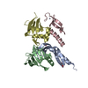
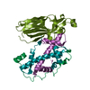
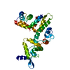
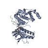
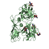
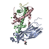
| Title | Crystal Structure of SltB3 from Pseudomonas aeruginosa in complex with NAG-NAM-pentapeptide | ||||||||||||
 Components Components | SOLUBLE LYTIC TRANGLYCOSILASE B3 | ||||||||||||
 Keywords Keywords | TRANSFERASE / CELL WALL RECYCLING | ||||||||||||
| Function / homology |  Function and homology information Function and homology informationpeptidoglycan lytic transglycosylase activity / peptidoglycan turnover / peptidoglycan catabolic process / metal ion binding Similarity search - Function | ||||||||||||
| Biological species |   PSEUDOMONAS AERUGINOSA (bacteria) PSEUDOMONAS AERUGINOSA (bacteria) | ||||||||||||
| Method |  X-RAY DIFFRACTION / SYNCHROTRON / MOLECULAR REPLACEMENT / Resolution: 2.23 Å X-RAY DIFFRACTION / SYNCHROTRON / MOLECULAR REPLACEMENT / Resolution: 2.23 Å | ||||||||||||
 Authors Authors | Dominguez-Gil, T. / Hermoso, J.A. | ||||||||||||
 Citation Citation | Journal: Acs Chem.Biol. / Year: 2016 Title: Turnover of Bacterial Cell Wall by Sltb3, a Multidomain Lytic Transglycosylase of Pseudomonas Aeruginosa. Authors: Lee, M. / Dominguez-Gil, T. / Hesek, D. / Mahasenan, K.V. / Lastochkin, E. / Hermoso, J.A. / Mobashery, S. | ||||||||||||
| History |
|
- Structure visualization
| Structure viewer | Molecule: MolmilJmol/JSmol |
|---|
- Downloads & links
-Download
| PDBx/mmCIF format | 5ao8.cif.gz | 148.5 KB | Display | PDBx/mmCIF format |
|---|---|---|---|---|
| PDB format | pdb5ao8.ent.gz | 117.1 KB | Display | PDB format |
| PDBx/mmJSON format | 5ao8.json.gz | Tree view | PDBx/mmJSON format | |
| Others |  Other downloads Other downloads |
-Validation report
| Arichive directory | https://data.pdbj.org/pub/pdb/validation_reports/ao/5ao8ftp://data.pdbj.org/pub/pdb/validation_reports/ao/5ao8 | HTTPS FTP |
|---|
-Related structure data
| Related structure data | 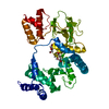 5anzSC 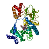 5ao7C S: Starting model for refinement C: citing same article ( |
|---|---|
| Similar structure data |


-Links
 PDBj
PDBj


- Assembly
| Deposited unit | 
| ||||||||
|---|---|---|---|---|---|---|---|---|---|
| 1 |
| ||||||||
| Unit cell |
|
-Components
| #1: Protein | Mass: 45034.430 Da / Num. of mol.: 1 / Fragment: UNP RESIDUES 33-448 Source method: isolated from a genetically manipulated source Source: (gene. exp.) PSEUDOMONAS AERUGINOSA (bacteria) / Production host: |
|---|---|
| #2: Polysaccharide | 2-acetamido-2-deoxy-beta-D-glucopyranose-(1-4)-methyl 2-acetamido-3-O-[(2R)-1-amino-1-oxopropan-2- ...2-acetamido-2-deoxy-beta-D-glucopyranose-(1-4)-methyl 2-acetamido-3-O-[(2R)-1-amino-1-oxopropan-2-yl]-2-deoxy-beta-D-glucopyranoside Type: oligosaccharide / Mass: 509.505 Da / Num. of mol.: 1 Source method: isolated from a genetically manipulated source |
| #3: Chemical | ChemComp-CA /   Mass: 40.078 Da / Num. of mol.: 1 / Source method: obtained synthetically / Formula: Ca Mass: 40.078 Da / Num. of mol.: 1 / Source method: obtained synthetically / Formula: Ca |
| #4: Sugar | ChemComp-NAG /   Type: D-saccharide, beta linking / Mass: 221.208 Da / Num. of mol.: 1 Type: D-saccharide, beta linking / Mass: 221.208 Da / Num. of mol.: 1Source method: isolated from a genetically manipulated source Formula: C8H15NO6 |
-Experimental details
-Experiment
| Experiment | Method: X-RAY DIFFRACTION |
|---|
- Sample preparation
| Crystal | Density Matthews: 1.8 Å3/Da / Density % sol: 31.75 % / Description: NONE |
|---|
-Data collection
| Diffraction | Mean temperature: 100 K |
|---|---|
| Diffraction source | Source: SYNCHROTRON / Site: ALBA  / Beamline: XALOC / Wavelength: 1 / Beamline: XALOC / Wavelength: 1 |
| Detector | Type: DECTRIS PILATUS 6M / Detector: PIXEL / Details: ELLIPTICALLY BENT MIRROR |
| Radiation | Monochromator: SI(111) CHANNEL-CUT, CRYOCOOLED / Protocol: SINGLE WAVELENGTH / Monochromatic (M) / Laue (L): M / Scattering type: x-ray |
| Radiation wavelength | Wavelength: 1 Å / Relative weight: 1 |
| Reflection | Resolution: 2.23→61 Å / Num. obs: 17322 / % possible obs: 99.9 % / Observed criterion σ(I): 0 / Redundancy: 8.5 % / CC1/2: 1 / Rmerge(I) obs: 0.01 / Net I/σ(I): 12.3 |
| Reflection shell | Resolution: 2.23→2.3 Å / Redundancy: 8.7 % / Rmerge(I) obs: 0.34 / Mean I/σ(I) obs: 4.2 / CC1/2: 0.98 / % possible all: 99.8 |
- Processing
| Software |
| |||||||||||||||||||||||||||||||||||||||||||||||||
|---|---|---|---|---|---|---|---|---|---|---|---|---|---|---|---|---|---|---|---|---|---|---|---|---|---|---|---|---|---|---|---|---|---|---|---|---|---|---|---|---|---|---|---|---|---|---|---|---|---|---|
| Refinement | Method to determine structure: MOLECULAR REPLACEMENT Starting model: PDB ENTRY 5ANZ Resolution: 2.23→53.766 Å / SU ML: 0.27 / σ(F): 1.34 / Phase error: 22.93 / Stereochemistry target values: ML
| |||||||||||||||||||||||||||||||||||||||||||||||||
| Solvent computation | Shrinkage radii: 0.9 Å / VDW probe radii: 1.11 Å / Solvent model: FLAT BULK SOLVENT MODEL | |||||||||||||||||||||||||||||||||||||||||||||||||
| Refinement step | Cycle: LAST / Resolution: 2.23→53.766 Å
| |||||||||||||||||||||||||||||||||||||||||||||||||
| Refine LS restraints |
| |||||||||||||||||||||||||||||||||||||||||||||||||
| LS refinement shell |
|
